Herunterladen Inhalt Inhalt Diese Seite drucken
Optika IM Serie Bedienungsanleitung
Vorschau ausblenden Andere Handbücher für IM Serie:
Inhaltsverzeichnis

Werbung

Verfügbare Sprachen
  • DE

Verfügbare Sprachen

  • DEUTSCH, seite 202
IM Series
INSTRUCTION MANUAL
Model
IM-3
IM-3F
IM-3FL4
IM-3LD2
IM-3LD4
IM-3LD4D
Ver. 5.2
2021

Werbung

Inhaltsverzeichnis
loading

Inhaltszusammenfassung für Optika IM Serie

  • Seite 1 IM Series INSTRUCTION MANUAL Model IM-3 IM-3F IM-3FL4 IM-3LD2 IM-3LD4 IM-3LD4D Ver. 5.2 2021...
  • Seite 2: Inhaltsverzeichnis

    Table of Contents Warning Symbols and conventions Safety Information Intended use Instrument description IM-3 IM-3F IM-3FL4 IM-3LD2 IM-3LD4 IM-3LD4D Unpacking IM-3 IM-3F / IM-3FL4 IM-3LD2 IM-3LD4 IM-3LD4D Assembling Installing the objectives Installing stage extension or mechanical stage Installing the stage insert Installing the eyepieces Installing the condenser Installing the LED housing...
  • Seite 3 14.5 Use of the Anti-glow cap 14.6 Filter holder / Shutter 14.6.1 Inserting a ND filter 14.6.2 Using the filter slider 15. Simultaneous observation in Phase Contrast + Fluorescence 15.1 IM-3F / IM-3FL4 15.2 IM-3LD2 / IM-3LD4 / IM-3LD4D 16. Microphotography 16.1 Use of C-mount cameras 16.2 Use of Reflex cameras 17. Maintenance 18.
  • Seite 4: Warning

    Warning This microscope is a scientific precision instrument designed to last for many years with a minimum of mainte- nance. It is built to high optical and mechanical standards and to withstand daily use. We remind you that this manual contains important information on safety and maintenance, and that it must therefore be made accessi- ble to the instrument users.
  • Seite 5: Instrument Description

    Instrument description IM-3 EYEPIECE HOUSING PHOTO/TV PORT FILTER HOLDER PHASE CONTRAST SLIDER CONDENSER DIOPTER STAGE ADJUSTMENT PLATE RING STAGE LIGHT PATH SELECTOR LEVER OBJECTIVE FINE FOCUS KNOB MAIN SWITCH MICROSCOPE BODY TENSION ADJUSTMENT COLLAR COARSE FOCUS KNOB Page 5...
  • Seite 6: Im-3F

    IM-3F PHOTO/TV PORT HOUSING EYEPIECE CONDENSER FILTER HOLDER PHASE CONTRAST SLIDER UV SHIELD DIOPTER ADJUSTMENT STAGE PLATE RING STAGE FLUORESCENCE LAMP HOUSING LIGHT PATH SELECTOR LEVER MICROSCOPE OBJECTIVE BODY FLUORESCENCE CUBE LEVER FLUORESCENCE MAIN SWITCH POWER SUPPLY FINE FOCUS KNOB TENSION ADJUSTMENT COLLAR...
  • Seite 7: Im-3Fl4

    IM-3FL4 HOUSING EYEPIECE CONDENSER PHOTO/TV PORT FILTER HOLDER DIOPTER ADJUSTMENT RING UV SHIELD STAGE PLATE LIGHT PATH STAGE SELECTOR LEVER FLUORESCENCE LAMP HOUSING FLUORESCENCE FLUORESCENCE CUBE LEVER POWER SUPPLY FINE FOCUS MICROSCOPE KNOB BODY TRANSMITTED LIGHT COARSE ADJUSTMENT KNOB FOCUS KNOB Page 7...
  • Seite 8: Im-3Ld2

    IM-3LD2 PHOTO/TV HOUSING PORT EYEPIECE CONDENSER DIOPTER ADJUSTMENT RING FILTER HOLDER PHASE CONTRAST SLIDER STAGE PLATE STAGE LIGHT PATH SELECTOR LEVER OBJECTIVE FLUORESCENCE REFLECTED LIGHT CUBE LEVER ADJUSTMENT KNOB FINE FOCUS KNOB TRANSMITTED LIGHT ADJUSTMENT KNOB MICROSCOPE BODY COARSE FOCUS KNOB Page 8...
  • Seite 9: Im-3Ld4

    IM-3LD4 PHOTO/TV HOUSING PORT EYEPIECE CONDENSER DIOPTER ADJUSTMENT RING FILTER HOLDER STAGE PLATE STAGE LIGHT PATH SELECTOR LEVER OBJECTIVE X-Y MOVEMENT KNOBS FLUORESCENCE REFLECTED LIGHT CUBE LEVER ADJUSTMENT KNOB FINE FOCUS KNOB TRANSMITTED LIGHT ADJUSTMENT KNOB MICROSCOPE BODY COARSE FOCUS KNOB Page 9...
  • Seite 10: Im-3Ld4D

    IM-3LD4D MONITOR CONDENSER FILTER HOLDER USB CAMERA EYEPIECE DIOPTER ADJUSTMENT RING STAGE PLATE STAGE LIGHT PATH SELECTOR LEVER OBJECTIVE X-Y MOVEMENT KNOBS FLUORESCENCE CUBE LEVER REFLECTED LIGHT ADJUSTMENT KNOB FINE FOCUS KNOB TRANSMITTED LIGHT ADJUSTMENT KNOB MICROSCOPE BODY COARSE FOCUS KNOB Page 10...
  • Seite 11 IM-3LD4D (back side) MONITOR POWER SUPPLY HDMI PLUG CONNECTOR MONITOR POWER POWER SUPPLY SUPPLY CABLE CABLE MOUSE / KEYBOARD RECEIVER PC STICK USB CABLE (TO CAMERA) PC STICK POWER SUPPLY CABLE HDMI CABLE (TO MONITOR) Page 11...
  • Seite 12: Unpacking

    Unpacking The microscope is housed in a moulded Styrofoam container. Remove the tape from the edge of the container and lift the top half of the container. Take some care to avoid that the optical items (objectives and eyepieces) fall out and get damaged. Using both hands (one around the arm and one around the base), lift the microscope from the container and put it on a stable desk.
  • Seite 13: Im-3F / Im-3Fl4

    IM-3F / IM-3FL4 ① ⑭ ② ⑮ ④ ⑨ ③ ⑩ ⑫ ⑬ ⑥ ⑤ ⑪ ⑯ ⑰ ⑱ ⑲ ⑦ ⑧ ① Microscope body ⑬ Fluorescence illuminator ② Condenser ⑭ HBO lamp housing ③ LED illuminator ⑮ Microscope power supply ④ Fluorescence power supply + cable ⑯ Phase contrast slider ⑤ Fluorescence power cable • P rovided with IM-3F ⑥ Filter holder • O ptional with IM-3FL4...
  • Seite 14: Im-3Ld2

    IM-3LD2 ③ ② ① ⑤ ⑭ ⑰ ⑦ ⑨ ⑯ ⑧ ⑩ ⑪ ④ ⑫ ⑮ ⑥ ⑬ ① Microscope body ⑩ Eyepieces ② Condenser ⑪ Stage inserts ③ LED illuminator ⑫ Centering telescope ④ Anti-glow cap ⑬ Green filter IF550 ⑤ Mechanical stage ⑭ Lens cleaning tissue ⑥ Phase contrast slider ⑮ UV shield ⑦ Glass insert for stage ⑯ Dust cover...
  • Seite 15: Im-3Ld4

    IM-3LD4 ③ ② ① ⑨ ⑫ ⑭ ⑦ ④ ⑬ ⑧ ⑪ ⑤ ⑥ ⑩ ① Microscope body ⑧ Metal insert for stage ② Condenser ⑨ Mechanical stage ③ LED illuminator ⑩ UV shield ④ Objectives ⑪ Stage inserts ⑤ Eyepieces ⑫ Lens cleaning tissue ⑥ Anti-glow cap ⑬ Dust cover ⑦ Glass insert for stage ⑭ Power supply + power cord Page 15...
  • Seite 16: Im-3Ld4D

    IM-3LD4D ③ ② ④ ⑨ ① ⑲ ⑪ ⑰ ⑦ ⑳ ⑧ ⑫ ⑤ ⑩ ⑥ ⑱ ⑬ ⑯ ⑭ ⑮ ① Microscope body ⑪ Camera ② Condenser ⑫ “C” mount ③ LED illuminator ⑬ Mouse ④ Anti-glow cap ⑭ Keyboard ⑤ Objectives ⑮ Monitor ⑥ Eyepieces ⑯ UV shield ⑦ Glass insert for stage...
  • Seite 17: Assembling

    Assembling Installing the objectives ② 1. Turn the coarse focusing knob ① until the nosepiece reaches its lowest position. • For a safe transport, the nosepiece is placed in the lowest position and the tension adjustment collar ② is adjusted to the proper tension when the mi- croscope leaves the factory.
  • Seite 18: Installing The Stage Insert

    Installing the stage insert 1. When using the glass stage, make sure that the insert is horizontal. 2. Install the stage insert in the stage opening. (Fig. 5) F ig. 5 ig. 5 Installing the eyepieces Insert both eyepieces into the tubes of the optical head. (Fig.
  • Seite 19: Installing The Led Housing

    Installing the LED housing • All models except IM-3LD4D 1. Insert the connector plug into the connector jack. 2. Push the LED housing gently into the holes of the illu- mination unit. (Fig. 9) F ig. 9 ig. 9 • IM-3LD4D The LED of the IM-3LD4D is already pre-installed on the illuminator.
  • Seite 20: Installing The Pc Screen (Im-3Ld4D)

    Installing the PC screen (IM-3LD4D) • For the camera installation, please refer to chapter 15.1. 1. Align the notch ① with the bracket ② on the illuminator. ① (Fig. 13) F ig. 13 ig. 13 ② 2. Once the monitor is fully inserted, tighten the wheel ③ to lock the monitor.
  • Seite 21: Connecting The Power Supply

    6. Plug the monitor power supply in the connector “F” on the monitor. (Fig. 17) 7. Plug the other side of the HDMI cable in the connector “G” on the monitor. (Fig. 17) F ig. 17 ig. 17 8. Plug the USB3.0 cable in the back side of the camera. (Fig.
  • Seite 22: Installing Fluorescence

    7.11 Installing fluorescence (IM-3F and IM3FL4 only) • Disconnect all electrical cables before installing or replacing the bulb. • The bulb has an anode and a cathode of different sizes. Respect the polarity during assembly, respecting the bulb dimensions. • Do not touch the bulb of the lamp with bare hands to leave no traces of grease on the bulb. If this happens, clean the bulb with a soft cloth before turning on the lamp.
  • Seite 23 4. Remove one of the knurled knobs from the filter holder and slide it into the slot placed in the back side of the microscope. (Fig. 22) 5. Once inserted, reinstall the knurled knob. F ig. 22 ig. 22 • IM-3F 6.
  • Seite 24 9. Open the lamp housing using the door lock screw ① and remove the lamp holder. (Fig. 26) ① F ig. 26 ig. 26 10. Remove the plastic block ② from the lamp holder (or the exhausted lamp in case of replacement) by loosening the two locking screws ③.
  • Seite 25 13. Connect the power cable to the external power supply. (Fig. 30) • The input voltage for fluorescence power supply is 110-240Vac. • Please use the standard power cable pro- vided. Select suitable one when missing or damaged. • Connect the power supply correctly, be sure to have a good earth connection.
  • Seite 26: Brightfield Observation (Transmitted Light)

    8. Brightfield observation (transmitted light) (Used commands) (Chapter) Main switch Set the main swith to “I” (ON) and adjust light intensity. Brightness adjustment knob Place a slide on the stage. Stage Insert 10x objective into the light path Nosepiece Coarse and fine focusing knobs Focus the specimen Observation head Adjust interpupillary distance...
  • Seite 27: Use Of The Microscope In Brightfield (Transmitted Light)

    Use of the microscope in brightfield (transmitted light) Turning on the microscope Move the main switch ① in the “I” (ON) position. (Fig. 31) ① F ig. 31 ig. 31 Adjusting the light intensity Turn the brightness adjustment knob ② placed on the left side of the microscope to increase and decrease the brightness.
  • Seite 28: Adjusting Interpupillary Distance

    Adjusting interpupillary distance ① Observing with both eyes, hold the two eyepiece prism as- semblies. Rotate them around their common axis until the fields of view match. • The graduation on the interpupillary distance indi- cator ①, pointed by the spot “.” on the eyepiece holder, shows the distance between the operator’s eyes.
  • Seite 29: Stage And Stage Inserts

    IM-3 LIGHT PATH LEVER BRIGHTNESS APPLICATION Binocular observation and microgra- 50% used for binocular observation / phy or video can be operated simul- 50% used for video or photography taneously 100% used for binocular observation / Binocular observation 0% used for video or photography IM-3F / IM-3FL4 / IM-3LD2 / IM-3LD4 / IM-3LD4D LIGHT PATH LEVER BRIGHTNESS...
  • Seite 30: Installing Stage Inserts

    9.8.1 Installing stage inserts 1. Install the holder in the mechanical stage. (Fig. 40) F F ig. 40 ig. 40 2. Multi well plates can be directly inserted in the mechan- ical stage. (Fig. 41) F ig. 41 ig. 41 Aperture diaphragm The Numerical Aperture (N.A.) value of the aperture diaphragm affects the image contrast.
  • Seite 31: Using Color Filters

    9.10 Using color filters Selecting the appropriate color filter according your need. (Fig. 44) FILTER Green (IF550) Phase contrast microscopy F ig. 44 ig. 44 Page 31...
  • Seite 32: Use Of The Microscope In Phase Contrast (Transmitted Light)

    10. Use of the microscope in phase contrast (transmitted light) 10.1 Installing the phase contrast slider 1. Insert the slider into the illumination system, printed face up. (Fig. 45) 2. Pull the slider into the desired position, until it arrives to the click stop.
  • Seite 33 4. Use the CT to focus the condenser phase ring (bright) ① and the objective phase ring (dark) ② image. If the bright phase ring’s image is not sharp, adjust the CT ① head until you can see a clear image of the phase ring. (Fig.
  • Seite 34: Use Of The Microscope In Rpc (Transmitted Light)

    11. Use of the microscope in RPC (transmitted light) Relief phase contrast (RPC) is a modification of conventional phase contrast that leads to visible improvements in image quality in optical microscopy. Specifically, the following parameters can be improved: contrast, focal depth, sharpness, three-dimensionality, flatness, and halo artifacts.
  • Seite 35: Rpc Observation

    11.3 RPC observation • RPC rings don’t need a centering. 1. Place a specimen on the stage and focus it. 2. Check that the RPC ring and the objective match, and that both are steadily set on a click stop. 3.
  • Seite 36: Fluorescence Observation (Im-3F / Im-3Fl4)

    12. Fluorescence observation (IM-3F / IM-3FL4) (Used commands) (Chapter) Set the power supply switch to “I” (ON) and Fluorescence power supply wait for the arc to stabilize (5 or 10 minutes). Place a specimen on the stage. Stage Fluorescence filter slider Insert in the light path the filtercube suitable for the specimen Nosepiece...
  • Seite 37: Use Of The Microscope In Fluorescence (Reflected Light)

    14. Use of the microscope in fluorescence (reflected light) 14.1 Centering the mercury bulb Only IM-3F / IM-3FL4 • Wait around 5 minutes before proceeding with this operation to allow the bulb to properly warm up. 1. Turn on the HBO mercury bulb by operating the power supply switch ①.
  • Seite 38 6. Using the centering screws ③ on the side of the lamp housing, center the image of the arc. (Fig. 59-60) F ig. 59 ig. 59 F ig. 60 ig. 60 7. Using the focusing screw of the collector lens ② enlarge the image until a homogeneous illumination is achieved.
  • Seite 39: Centering The Field Diaphragm

    14.2 Centering the field diaphragm ② ① Only IM-3F / IM-3FL4 1. Place the specimen on the stage, insert 10x objective into the light path and focus. 2. Rotate the field diaphragm lever ①, to fully close the diaphragm. (Fig. 63) 3. Rotate the two centering screws ② to bring the bright spot in the center of the field of view.
  • Seite 40: Available Fluorescence Filter Cubes

    • IM-3FL4 Move the filter lever (placed on the left side of the microscope) ⑤ to insert the desired filter cube: B, G (V and UV - optional). (Fig. 67) ⑤ F ig. 67 ig. 67 • IM-3LD2 / IM-3LD4 / IM-3LD4D Move the filter lever (placed on the left side of the microscope) ⑥...
  • Seite 41: Installing Fluorescence Filter

    • IM-3LD4 / IM-3LD4D F I LT E R EXCITATION D I C H R O I C BARRIER APPLICATIONS NAME FILTER MIRROR FILTER • DAPI M-1233 325-375 nm 415 nm 435LP nm • Hoechst • Coumarin M-1232 390-420 nm 440 nm 450LP nm •...
  • Seite 42: Use Of The Anti-Glow Cap

    6. Reinstall the filter slider on the microscope. 7. Close the side cover. 8. Apply the adhesive marker ⑦ for the fluorescence cube on the side cover. (Fig. 71) 9. Connect the power supply. 10. Start working. ⑦ F ig. 71 ig.
  • Seite 43: Using The Filter Slider

    14.6.2 Using the filter slider • Moving the slider toward left or right to insert the desired position. A click sound will confirm the proper positioning of the slider. 1. When the position ① is inserted (provided that a ND filter is installed) the light coming from the lamp housing will be decreased, according to the filter type installed.
  • Seite 44: Simultaneous Observation In Phase Contrast + Fluorescence

    15. Simultaneous observation in Phase Contrast + Fluorescence • Fluorescence models allow observation in transmitted light Phase contrast in combination with reflected light Fluorescence. Samples with rapid decay must first be observed in Fluorescence and then in Phase Contrast. The combined observation allows you to easily identify some areas of the sample that emit fluorescence. 15.1 IM-3F / IM-3FL4 1.
  • Seite 45: Microphotography

    16. Microphotography 16.1 Use of C-mount cameras ② 1. Loosen the clamping screw ① on the trinocular port and remove the dust cap ②. (Fig. 75) ① F ig. 75 ig. 75 2. Screw the C-mount adapter ③ to the camera ④ and insert the round dovetail of the C-mount into the emp- ty hole of the trinocular port, then tighten the clamping ④...
  • Seite 46: Maintenance

    Do not disassemble objectives or eyepieces in attempt to clean them. For the best results, use the OPTIKA cleaning kit (see catalogue). If you need to send the microscope to Optika for maintenance, please use the original packaging. Page 46...
  • Seite 47: Troubleshooting

    18. Troubleshooting Review the information in the table below to troubleshoot operating problems. PROBLEM CAUSE SOLUTION I. Optical Section: The illumination is on, but the field of The plug of the LED holder is not con- Connect them view is dark. nected to the illumination set The brightness is too low Adjust to a proper setting...
  • Seite 48 III. Electric section The LED doesn’t turn on. No power supply Check the power cord connection The brightness is not enough The brightness adjustment is low Adjust the brightness The light blinks The power cord is poorly connected Check the power cord IV.
  • Seite 49: Equipment Disposal

    Equipment disposal Art.13 Dlsg 25 July 2005 N°151. “According to directives 2002/95/EC, 2002/96/EC and 2003/108/EC relating to the reduction in the use of hazardous substances in electrical and electronic equipment and waste disposal.” The basket symbol on equipment or on its box indicates that the product at the end of its useful life should be collected separately from other waste.
  • Seite 50 OPTIKA S.r.l. ® Via Rigla, 30 - 24010 Ponteranica (BG) - ITALY Tel.: +39 035.571.392 info@optikamicroscopes.com - www.optikamicroscopes.com OPTIKA Spain spain@optikamicroscopes.com OPTIKA USA usa@optikamicroscopes.com OPTIKA China china@optikamicroscopes.com OPTIKA India india@optikamicroscopes.com OPTIKA Central America camerica@optikamicroscopes.com...
  • Seite 51 Serie IM MANUALE DI ISTRUZIONI Modello IM-3 IM-3F IM-3FL4 IM-3LD2 IM-3LD4 IM-3LD4D Ver. 5.2 2021...
  • Seite 52 Sommario Avvertenza Simboli Informazioni sulla sicurezza Utilizzo previsto Descrizione dello strumento IM-3 IM-3F IM-3FL4 IM-3LD2 IM-3LD4 IM-3LD4D Disimballaggio IM-3 IM-3F / IM-3FL4 IM-3LD2 IM-3LD4 IM-3LD4D Assemblaggio Montaggio degli obiettivi Montaggio di estensione laterale e traslatore Montaggio del piattello Montaggio degli oculari Montaggio del condensatore Montaggio dell’alloggiamento LED 7.7 Montaggio dei filtri colorati...
  • Seite 53 14.5 Uso del tappo anti-riflesso 14.6 Portafiltri / Shutter 14.6.1 Inserire un filtro ND 14.6.2 Uso della slitta portafiltri 15. Osservazione simultanea in Contrasto di Fase + Fluorescenza 15.1 IM-3F / IM-3FL4 15.2 IM-3LD2 / IM-3LD4 / IM-3LD4D 16. Microfotografia 16.1 Uso di telecamere a passo “C” 16.2 Uso di fotocamere Reflex 17. Manutenzione 18. Risoluzione dei problemi Smaltimento Pagina 53...
  • Seite 54: Shock Elettrico

    Avvertenza Questo microscopio è uno strumento scientifico di alta precisione, progettato per durare a lungo con una minima manutenzione; la realizzazione è secondo i migliori standard ottici e meccanici, per poter essere utilizzato quotidianamente. Vi ricordiamo che questo manuale contiene informazioni importanti per la sicurezza e per la manutenzione dello strumento, e deve quindi essere messo a disposizione di coloro che lo utilizzeranno.
  • Seite 55: Descrizione Dello Strumento

    Descrizione dello strumento IM-3 OCULARI ALLOGGIAMENTO USCITA FOTO/TV PORTA-FILTRI SLITTA PER CONTRASTO DI FASE CONDENSATORE ANELLO DI REGOLAZIONE PIATTELLO DIOTTRICA TAVOLINO SELETTORE PERCORSO OTTICO OBIETTIVI MANOPOLA MICROMETRICA INTERRUTTORE STATIVO MICROSCOPIO ANELLO DI REGOLAZIONE DELLA TENSIONE MANOPOLA MACROMETRICA Pagina 55...
  • Seite 56 IM-3F USCITA FOTO/TV ALLOGGIAMENTO OCULARI CONDENSATORE PORTA-FILTRI SLITTA PER CONTRASTO DI FASE SCHERMO UV ANELLO DI REGOLAZIONE PIATTELLO DIOTTRICA TAVOLINO CORPO LAMPADA FLUORESCENZA SELETTORE PERCORSO OTTICO STATIVO OBIETTIVI MICROSCOPIO SELETTORE CUBI FLUORESCENZA ALIMENTATORE INTERRUTTORE FLUORESCENZA MANOPOLA ANELLO DI MICROMETRICA REGOLAZIONE DELLA TENSIONE MANOPOLA MACROMETRICA...
  • Seite 57 IM-3FL4 ALLOGGIAMENTO OCULARI CONDENSATORE USCITA FOTO/TV PORTA-FILTRI ANELLO DI REGOLAZIONE DIOTTRICA SCHERMO UV PIATTELLO SELETTORE TAVOLINO PERCORSO OTTICO CORPO LAMPADA FLUORESCENZA SELETTORE CUBI ALIMENTATORE FLUORESCENZA FLUORESCENZA MANOPOLA MICROMETRICA STATIVO MICROSCOPIO MANOPOLA MANOPOLA REGOLAZIONE MACROMETRICA LUCE TRASMESSA Pagina 57...
  • Seite 58 IM-3LD2 ALLOGGIAMENTO USCITA FOTO/TV OCULARI CONDENSATORE ANELLO DI REGOLAZIONE DIOTTRICA PORTA-FILTRI SLITTA PER CONTRASTO DI FASE PIATTELLO TAVOLINO SELETTORE PERCORSO OTTICO OBIETTIVI SELETTORE CUBI MANOPOLA FLUORESCENZA REGOLAZIONE LUCE RIFLESSA MANOPOLA MICROMETRICA MANOPOLA REGOLAZIONE LUCE TRASMESSA STATIVO MICROSCOPIO MANOPOLA MACROMETRICA Pagina 58...
  • Seite 59 IM-3LD4 ALLOGGIAMENTO USCITA FOTO/TV OCULARI CONDENSATORE ANELLO DI REGOLAZIONE DIOTTRICA PORTA-FILTRI PIATTELLO TAVOLINO SELETTORE PERCORSO OTTICO OBIETTIVI MANOPOLE TRASLAZIONE SELETTORE CUBI MANOPOLA FLUORESCENZA REGOLAZIONE LUCE RIFLESSA MANOPOLA MICROMETRICA MANOPOLA REGOLAZIONE LUCE TRASMESSA STATIVO MICROSCOPIO MANOPOLA MACROMETRICA Pagina 59...
  • Seite 60 IM-3LD4D MONITOR CONDENSATORE PORTA-FILTRI TELECAMERA USB OCULARI ANELLO DI REGOLAZIONE DIOTTRICA PIATTELLO TAVOLINO SELETTORE PERCORSO OTTICO OBIETTIVI MANOPOLE TRASLAZIONE SELETTORE CUBI FLUORESCENZA MANOPOLA REGOLAZIONE LUCE RIFLESSA MANOPOLA MICROMETRICA MANOPOLA REGOLAZIONE STATIVO LUCE TRASMESSA MICROSCOPIO MANOPOLA MACROMETRICA Pagina 60...
  • Seite 61 IM-3LD4D (retro) CONNETTORE ALIMENTAZIONE CONNETTORE HDMI MONITOR CAVO CAVO ALIMENTAZIONE ALIMENTAZIONE MONITOR RICEVITORE MOUSE / TASTIERA USB PC STICK CAVO USB (ALLA TELECAMERA) CAVO ALIMENTAZIONE PC STICK CAVO HDMI (AL MONITOR) Pagina 61...
  • Seite 62: Disimballaggio

    Disimballaggio Il microscopio è riposto in un imballo di polistirolo espanso. Rimuovere il nastro adesivo dal collo ed aprire la parte superiore dell’imballo. Fare attenzione a non far cadere le parti ottiche (obiettivi e oculari) nell’estrarre il microscopio dalla scatola per evitare che vengano danneggiati. Utilizzare entrambe le mani (una intorno allo stativo e una alla base), sfilare il microscopio dal contenitore e appoggiarlo su un piano stabile.
  • Seite 63: Im-3F / Im-3Fl4

    IM-3F / IM-3FL4 ① ⑭ ② ⑮ ④ ⑨ ③ ⑩ ⑫ ⑬ ⑥ ⑤ ⑪ ⑯ ⑰ ⑱ ⑲ ⑦ ⑧ ① Stativo del microscopio ⑬ Illuminatore per fluorescenza ② Condensatore ⑭ Corpo lampada HBO ③ Alloggiamento LED ⑮ Alimentatore microscopio ④ Alimentatore per fluorescenza + cavo ⑯ Slitta contrasto di fase ⑤ Cavo di connessione fluorescenza • In dotazione con IM-3F ⑥ Porta-filtri • Opzionale con IM-3FL4...
  • Seite 64: Im-3Ld2

    IM-3LD2 ③ ② ① ⑤ ⑭ ⑰ ⑦ ⑨ ⑯ ⑧ ⑩ ⑪ ④ ⑫ ⑮ ⑥ ⑬ ① Stativo del microscopio ⑩ Oculari ② Condensatore ⑪ Inserti per tavolino ③ Alloggiamento LED ⑫ Telescopio di centramento ⑬ Filtro verde IF550 ④ Tappo anti riflesso ⑤ Traslatore ⑭ Cartine pulizia ottica ⑥ Slitta per contrasto di fase ⑮ Schermo UV ⑦ Disco in vetro ⑯ Copertina antipolvere...
  • Seite 65: Im-3Ld4

    IM-3LD4 ③ ② ① ⑨ ⑫ ⑭ ⑦ ④ ⑬ ⑧ ⑪ ⑤ ⑥ ⑩ ① Stativo del microscopio ⑧ Disco in metallo ② Condensatore ⑨ Traslatore ③ Alloggiamento LED ⑩ Schermo UV ④ Obiettivi ⑪ Inserti per tavolino ⑤ Oculari ⑫ Cartine pulizia ottica ⑥ Tappo anti riflesso ⑬ Copertina antipolvere ⑦ Disco in vetro ⑭ Alimentatore + cavo Pagina 65...
  • Seite 66: Im-3Ld4D

    IM-3LD4D ③ ② ④ ⑨ ① ⑲ ⑪ ⑰ ⑦ ⑳ ⑧ ⑫ ⑤ ⑩ ⑥ ⑱ ⑬ ⑯ ⑭ ⑮ ① Stativo del microscopio ⑪ Telecamera ② Condensatore ⑫ Passo “C” ③ Alloggiamento LED ⑬ Mouse ⑭ Tastiera ④ Tappo anti riflesso ⑤ Obiettivi ⑮ Monitor ⑥ Oculari ⑯ Schermo UV ⑦ Disco in vetro...
  • Seite 67: Assemblaggio

    Assemblaggio Montaggio degli obiettivi ② 1. Ruotare la manopola di regolazione macrometrica ① finché la torretta portaobiettivi si trova nella posizione più bassa. • Per garantire la sicurezza durante il trasporto, pri- ma della spedizione la torretta viene messa nella posizione più...
  • Seite 68: Montaggio Del Piattello

    Montaggio del piattello 1. Assicurarsi che il piano portapreparati sia perfettamen- te orizzontale quando si usa il piano in vetro. 2. Inserire l’inserto nell’apertura del piano. (Fig. 5) F ig. 5 ig. 5 Montaggio degli oculari Togliere il tappo ai tubi portaoculari, inserire gli oculari nei tubi.
  • Seite 69: Montaggio Dell'alloggiamento Led

    Montaggio dell’alloggiamento LED • Tutti i modelli tranne IM-3LD4D 1. Inserire il cavo di alimentazione nel connettore jack. 2. Inserire il portalampada spingendolo con molta cura nei fori del gruppo di illuminazione. (Fig. 9) F ig. 9 ig. 9 • IM-3LD4D Il LED dell’IM-3LD4D è...
  • Seite 70: Montaggio Del Monitor Pc (Im-3Ld4D)

    Montaggio del monitor PC (IM-3LD4D) • Per l’installazione della telecamera, fare riferimento al capitolo 15.1. 1. Allineare la tacca ① con la staffa ② sull’illuminatore. ① (Fig. 13) F ig. 13 ig. 13 ② 2. Una volta che il monitor è completamente inserito, ser- rare la rotella ③...
  • Seite 71: Connettere L'alimentatore

    6. Collegare l’alimentatore del monitor nel connettore “F” sul monitor. (Fig. 17) 7. Inserire l’altro lato del cavo HDMI nel connettore “G” del monitor. (Fig. 17) F ig. 17 ig. 17 8. Collegare il cavo USB3.0 nel retro della telecamera. (Fig.
  • Seite 72: Montaggio Della Fluorescenza

    7.11 Montaggio della fluorescenza (Solo IM-3F e IM-3FL4) • Disconnettere tutti i cavi elettrici prima di procedere all’installazione o alla sostituzione della lampada. • La lampada ha un anodo ed un catodo con dimensioni diverse. Rispettare le polarità in fase di montaggio, rispettando le dimensioni di attacco della lampada. •...
  • Seite 73 4. Rimuovere una delle manopole zigrinate dalla slitta portafiltri e inserire la slitta nella fessura posta sul retro del microscopio. (Fig. 22) 5. Una volta inserita la slitta, riavvitare la manopola zigrinata. F ig. 22 ig. 22 • IM-3F 6. Avvitare sull’estremità dell’astina il terminale con la scritta G incisa.
  • Seite 74 9. Aprire il corpo lampada usando la vite di serraggio dello sportello ① ed estrarre il supporto lampada. (Fig. 26) ① F ig. 26 ig. 26 10. Rimuovere il blocco in plastica ② dal corpo lampada (o la lampada esausta in caso di sostituzione) allentando le due viti di bloccaggio ③.
  • Seite 75 13. Inserire il cavo di alimentazione nel connettore. (Fig. • La tensione di ingresso dell’alimentatore per fluorescenza è 110-240Vac. • Si prega di utilizzare il cavo di alimenta- zione standard fornito. Selezionare un cavo idoneo in caso di cavo mancante o danneggiato. • Collegare l’alimentatore in modo corret- to, assicurandosi di avere una buona messa a terra.
  • Seite 76: Osservazione In Campo Chiaro (Luce Trasmessa)

    Osservazione in campo chiaro (luce trasmessa) (Comandi utilizzati) (Capitolo) Portare su “I” (ON) l’interruttore generale e regolare Interruttore generale l’intensità luminosa. Selettore regolazione intensità Porre un preparato sul tavolino. Tavolino Revolver Inserire l’obiettivo 10x nel percorso ottico Manopole macro e micrometrica di mes- Mettere a fuoco il preparato sa a fuoco Tubo binoculare...
  • Seite 77: Uso Del Microscopio In Campo Chiaro (Luce Trasmessa)

    Uso del microscopio in campo chiaro (luce trasmessa) Accensione del microscopio Spostare l’interruttore principale ① in posizione “I” (ON). (Fig. 31) ① F ig. 31 ig. 31 Regolazione dell’intensità luminosa Agire sulla rotellina di regolazione dell’intensità luminosa ② per aumentare o diminuire il voltaggio dell’illuminazione. (Fig.
  • Seite 78: Regolazione Della Distanza Interpupillare

    Regolazione della distanza interpupillare ① Osservando con entrambi gli occhi, sostenere il gruppo di oculari. Ruotare questi lungo l’asse comune fino ad ottenere un unico campo visivo. • La scala graduata sull’indicatore della distanza interpupillare ①, indicata dal puntino “.” sul portaoculare, mostra la distanza interpupillare dell’operatore.
  • Seite 79: Traslatore E Portapreparati

    IM-3 LEVA SELETTORE LUMINOSITÀ APPLICAZIONI PERCORSO OTTICO Osservazione binoculare e micro- 20% per osservazione binoculare / 80% fotografia possono essere eseguite per microfotografia simultaneamente 100% per osservazione binoculare / 0% Osservazione binoculare per microfotografia IM-3F / IM-3FL4 / IM-3LD2 / IM-3LD4 / IM-3LD4D LEVA SELETTORE LUMINOSITÀ...
  • Seite 80: Montaggio Degli Inserti

    9.8.1 Montaggio degli inserti 1. Montare il supporto nel traslatore. (Fig. 40) F F ig. 40 ig. 40 2. Le piastre a pozzetti multipli possono essere inserite direttamente nel traslatore. (Fig. 41) F ig. 41 ig. 41 Diaframma di apertura Il valore di apertura numerica (A.N.) del diaframma di apertura influenza il contrasto dell’immagine.
  • Seite 81: Uso Dei Filtri Colorati

    9.10 Uso dei filtri colorati Selezionare il filtro colorato adatto alle proprie necessità. (Fig. 44) FILTRO Verde Microscopia a contrasto di fase F ig. 44 ig. 44 Pagina 81...
  • Seite 82: Uso Del Microscopio In Contrasto Di Fase (Luce Trasmessa)

    10. Uso del microscopio in contrasto di fase (luce trasmessa) 10.1 Montaggio della slitta per contrasto di fase 1. Inserire la slitta nel gruppo illuminatore, con la parte stampata verso l’alto. (Fig. 45) 2. Muovere la slitta nella posizione desiderata finché con si blocca con un click.
  • Seite 83 4. Con il CT mettere a fuoco l’immagine dell’anello di fase del condensatore (chiaro) ① e dell’obiettivo (scuro) ②. ① Se l’immagine dell’anello chiaro non è nitida, regolare il CT fino ad ottenere un’immagine nitida dell’anello. (Fig. 5. Regolare le viti dei due fori di centratura sulla slitta per contrasto di fase con le brugole in dotazione fino a far coincidere l’anello chiaro con l’anello scuro.
  • Seite 84: Uso Del Microscopio In Rpc (Luce Trasmessa)

    11. Uso del microscopio in RPC (luce trasmessa) Il contrasto di fase a rilievo (RPC) è una modifica del contrasto di fase convenzionale che porta a miglioramenti visibili della qualità dell’immagine nella microscopia ottica. In particolare, i seguenti parametri possono essere migliorati: contrasto, profondità...
  • Seite 85: Osservazione In Rpc

    11.3 Osservazione in RPC • Gli anelli RPC non hanno bisogno di centraggio. 1. Posizionare un preparato sul piano e metterlo a fuoco. 2. Verificare che l’anello RPC e l’obiettivo corrispondano e che entrambi siano fissi in posizione di blocco. 3.
  • Seite 86: Osservazione In Fluorescenza (Im-3F / Im-3Fl4)

    12. Osservazione in fluorescenza (IM-3F / IM-3FL4) (Comandi utilizzati) (Capitolo) Portare su “I” (ON) l’interruttore generale ed attendere che Alimentatore fluorescenza l’arco si stabilizzi (5 o 10 minuti). Porre un preparato sul tavolino. Tavolino Inserire nel percorso ottico il filtro per fluorescenza Selettore filtri fluorescenza adatto al campione Revolver Inserire nel percorso ottico l’obiettivo desiderato e...
  • Seite 87: Uso Del Microscopio In Fluorescenza (Luce Riflessa)

    14. Uso del microscopio in fluorescenza (luce riflessa) 14.1 Centraggio della lampada HBO Solo IM-3F / IM-3FL4 • Attendere circa 5 minuti prima di procedere a questa operazione per permettere alla lampada di riscaldarsi correttamente. 1. Accendere la lampada a vapori di mercurio agendo sull’interruttore dell’alimentatore ①. (Fig. 55) ①...
  • Seite 88 6. Usando le viti di centraggio ③ poste sul lato del corpo lampada centrare l’immagine dell’arco. (Fig. 59-60) F ig. 59 ig. 59 F ig. 60 ig. 60 7. Usando la vite di messa a fuoco della lente collettrice ② allargare l’immagine fino ad ottenere un’illuminazione omogenea.
  • Seite 89: Centraggio Del Diaframma Di Campo

    14.2 Centraggio del diaframma di campo ① ② Solo IM-3F / IM-3FL4 1. Posizionare il campione sul tavolino, inserire l’obiettivo 10x nel percorso ottico e mettere a fuoco. 2. Ruotare la leva del diaframma di campo ①, per chiudere completamente il diaframma. (Fig. 63) 3.
  • Seite 90: Filtri Per Fluorescenza Disponibili

    • IM-3FL4 Spostare la leva del selettore (posta sulla sinistra del microscopio) ⑤ per inserire il filtro desiderato: B, G (V e UV - opzionali). (Fig. 67) ⑤ F ig. 67 ig. 67 • IM-3LD2 / IM-3LD4 / IM-3LD4D Spostare la leva del selettore (posta sulla sinistra del microscopio) ⑥...
  • Seite 91: Montaggio Dei Filtri Per Fluorescenza

    • IM-3LD4 / IM-3LD4D CUBO FILTRO DI SPECCHIO FILTRO DI APPLICAZIONI FILTRO ECCITAZIONE DICROICO EMISSIONE • DAPI M-1233 325-375 nm 415 nm 435LP nm • Hoechst • Coumarin M-1232 390-420 nm 440 nm 450LP nm • Pacific Blue • FITC M-1230 455-495 nm 500 nm...
  • Seite 92: Uso Del Tappo Anti-Riflesso

    6. Rimontare la slitta filtri sul microscopio. 7. Chiudere il coperchio laterale. 8. Applicare il marcatore adesivo ⑦ per il cubo per fluore- scenza sul coperchio laterale. (Fig. 71) 9. Collegare l’alimentatore. 10. Iniziare a lavorare. ⑦ F ig. 71 ig.
  • Seite 93: Uso Della Slitta Portafiltri

    14.6.2 Uso della slitta portafiltri • Spostare la slitta verso destra o verso sinistra per inserire la posizione desiderata. Uno scattino confermerà la corretta posizione della slitta. 1. Inserendo la posizione ① (a condizione che sia installato un filtro ND) la luce proveniente dalla lampada verrà ridotta in funzione del tipo di filtro installato.
  • Seite 94: Osservazione Simultanea In Contrasto Di Fase + Fluorescenza

    15. Osservazione simultanea in Contrasto di Fase + Fluorescenza • I modelli a fluorescenza consentono l’osservazione in luce trasmessa Contrasto di Fase in combinazione con la Fluorescenza in luce riflessa. I campioni con decadimento rapido devono essere osservati prima in Fluorescenza e quindi in Contrasto di Fase. L’osservazione combinata consente di identificare facilmente alcune aree del campione che emettono fluorescenza. 15.1 IM-3F / IM-3FL4 1. Accendere l’alimentatore per la lampada a fluorescenza HBO ed attendere 5 minuti prima che l’arco si stabilizzi.
  • Seite 95: Microfotografia

    16. Microfotografia 16.1 Uso di telecamere a passo “C” ② 1. Allentare la vite di bloccaggio ① sul tubo trinoculare e rimuovere il tappo antipolvere ②. (Fig. 75) ① F ig. 75 ig. 75 2. Avvitare l’adattatore passo C ③ alla telecamera ④ e installare l’attacco rotondo del passo C nel foro vuoto del tubo trinoculare, quindi riavvitare la vite di serraggio ①.
  • Seite 96: Manutenzione

    Non smontare gli obiettivi o gli oculari per cercare di pulirli. Per un migliore risultato, utilizzare il kit di pulizia OPTIKA (vedi catalogo). Se si necessita di spedire il microscopio al produttore per la manutenzione, si prega di utilizzare l’imballo originale.
  • Seite 97: Risoluzione Dei Problemi

    18. Risoluzione dei problemi Consultare le informazioni riportate nella tabella sottostante per risolvere eventuali problemi operativi. PROBLEMA CAUSA SOLUZIONE I. Sezione Ottica: L’illuminatore è acceso, ma il campo La spina dell’alloggiamento LED non è Collegare l’alloggiamento LED al grup- visivo è scuro collegata al gruppo illuminatore po illuminatore La luminosità...
  • Seite 98 II. Sezione Meccanica La manopola macrometrica è difficile La manopola macrometrica è difficile La manopola macrometrica è difficile da ruotare da ruotare da ruotare La messa a fuoco è instabile La frizione della messa a fuoco è rego- Stringere la frizione lata bassa III.
  • Seite 99: Smaltimento

    Smaltimento Ai sensi dell’articolo 13 del decreto legislativo 25 luglio 2005 n°151. “Attuazione delle direttive 2002/95/CE, 2002/96/CE e 2003/108/CE, relative alla riduzione dell’uso di sostanze pericolose nelle apparecchiature elettri- che ed elettroniche, nonché allo smaltimento dei rifiuti”. Il simbolo del cassonetto riportato sulla apparecchiatura o sulla sua confezione indica che il prodotto alla fine della propria vita utile deve essere raccolto separatamente degli altri rifiuti.
  • Seite 100 OPTIKA S.r.l. ® Via Rigla, 30 - 24010 Ponteranica (BG) - ITALY Tel.: +39 035.571.392 info@optikamicroscopes.com - www.optikamicroscopes.com OPTIKA Spain spain@optikamicroscopes.com OPTIKA USA usa@optikamicroscopes.com OPTIKA China china@optikamicroscopes.com OPTIKA India india@optikamicroscopes.com OPTIKA Central America camerica@optikamicroscopes.com...
  • Seite 101 Serie IM MANUAL DE INSTRUCCIONES Modelo IM-3 IM-3F IM-3FL4 IM-3LD2 IM-3LD4 IM-3LD4D Ver. 5.2 2021...
  • Seite 102 Indice Advertencia Símbolos Información de seguridad Utilización Descripción del instrumento IM-3 IM-3F IM-3FL4 IM-3LD2 IM-3LD4 IM-3LD4D Desembalaje IM-3 IM-3F / IM-3FL4 IM-3LD2 IM-3LD4 IM-3LD4D Montaje Montaje de los objetivos Montaje de extensión lateral y del carro móvil Instalación del disco Instalación de los oculares Instalación del condensador Instalación del LED...
  • Seite 103 14.5 Uso de la tapa antirreflejo 14.6 Portafiltros / Obturador 14.6.1 Insertar un filtro ND 14.6.2 Uso del la corredera del filtro 15. Observación simultánea Contraste de fase + Fluorescencia 15.1 IM-3F / IM-3FL4 15.2 IM-3LD2 / IM-3LD4 / IM-3LD4D 16. Microfotografía 16.1 Uso de cámaras de paso “C” 16.2 Uso de cámara Reflex 17. Mantenimiento 18. Guía de solución de problemas Medidas ecológicas y reciclaje Página 103...
  • Seite 104: Símbolos

    Advertencia Este microscopio es un instrumento científico de precisión. Su utilización está pensada para una larga duración con un mínimo nivel de mantenimiento. Para su fabricación se han utilizado elementos ópticos y mecánicos de elevada calidad que lo convierten en el instrumento ideal para la utilización diaria en las aulas y el laboratorio. Informamos que esta guía contiene importantes informaciones sobre la seguridad y el mantenimiento del pro- ducto y por lo tanto debe ser accesible a todos aquellos que utilizan dicho instrumento.
  • Seite 105: Descripción Del Instrumento

    Descripción del instrumento IM-3 OCULARE SOPORTE SALIDA FOTO/ MONTURA PARA FILTROS CORREDERA DE CON- TRASTE DE FASE CONDENSADOR ANILLO DE REGULACIÓN PLACA DIÓPTRICA PLATINA PALANCA DE SELECCIÓN DEL CAMINO DE LUZ OBJETIVOS MANDO DEL ENFOQUE MACROMÉTRICO INTERRUPTOR PRIN- CIPAL CUERPO DEL MICROSCOPIO ANILLO DE REGULACIÓN DE...
  • Seite 106: Im-3F

    IM-3F SALIDA FOTO/ SOPORTE OCULAR CONDENSADOR MONTURA PARA FILTROS CORREDERA DE CONTRASTE DE FASE PANTALLA UV ANILLO DE REGULACIÓN PLACA DIÓPTRICA PLATINA CUERPO LÁMPARA FLUORESCENTE PALANCA DE SELECCIÓN DEL CAMINO DE LUZ CUERPO DEL OBJETIVOS MICROSCOPIO PALANCA DEL CUBO DE FLUORESCENCIA INTERRUPTOR PRIN- ALIMENTACIÓN DE...
  • Seite 107: Im-3Fl4

    IM-3FL4 SOPORTE OCULAR CONDENSADOR SALIDA FOTO/TV MONTURA PARA FILTROS ANILLO DE REGULACIÓN DIÓPTRICA PANTALLA UV PLACA PALANCA DE PLATINA SELECCIÓN DEL CAMINO DE LUZ CUERPO LÁMPARA FLUORESCENTE PALANCA DEL ALIMENTACIÓN DE CUBO DE FLUORESCENCIA FLUORESCENCIA MANDO DEL ENFOQUE MICROMÉTRICO CUERPO DEL MICROSCOPIO PERILLA DE AJUSTE DE MANDO DEL ENFOQUE...
  • Seite 108: Im-3Ld2

    IM-3LD2 SOPORTE SALIDA FOTO/ OCULAR CONDENSADOR ANILLO DE REGULACIÓN DIÓPTRICA MONTURA PARA FILTROS CORREDERA DE CONTRASTE DE FASE PLACA PLATINA PALANCA DE SELECCIÓN DEL CAMINO DE LUZ OBJETIVOS PALANCA DEL PERILLA DE AJUSTE DE CUBO DE LA LUZ REFLEJADA FLUORESCENCIA MANDO DEL ENFOQUE MICROMÉTRICO PERILLA DE AJUSTE DE...
  • Seite 109: Im-3Ld4

    IM-3LD4 SOPORTE SALIDA FOTO/TV OCULAR CONDENSADOR ANILLO DE REGULACIÓN DIÓPTRICA MONTURA PARA FILTROS PLACA PLATINA PALANCA DE SELECCIÓN DEL CAMINO DE LUZ OBJETIVOS PERILLAS DE MOVIMIENTO PALANCA DEL CUBO DE PERILLA DE AJUSTE DE FLUORESCENCIA LA LUZ REFLEJADA MANDO DEL ENFOQUE MICROMÉTRICO PERILLA DE AJUSTE DE LA LUZ TRANSMITIDA...
  • Seite 110: Im-3Ld4D

    IM-3LD4D MONITOR CONDENSADOR MONTURA PARA CÁMARA USB FILTROS OCULAR ANILLO DE REGULACIÓN DIÓPTRICA PLACA PLATINA PALANCA DE SELECCIÓN DEL CAMINO DE LUZ OBJETIVOS PERILLAS DE MOVIMIENTO PALANCA DEL CUBO DE FLUORESCENCIA PERILLA DE AJUSTE DE LA LUZ REFLEJADA MANDO DEL ENFOQUE MICROMÉTRICO PERILLA DE AJUSTE DE LA LUZ TRANSMITIDA...
  • Seite 111 IM-3LD4D (lado posterior) CONECTOR DE ALIMENTACIÓN DEL MONITOR CONECTOR HDMI CABLE DE ALI- CABLE DE MENTACIÓN DEL ALIMENTACIÓN DEL MONITOR RECEPTOR USB RATÓN / TECLADO PC STICK CABLE USB (A LA CÁMARA) CABLE DE ALIMENTACIÓN DEL PC STICK CABLE HDMI (AL MONITOR) Página 111...
  • Seite 112: Desembalaje

    Desembalaje El microscopio esta embalado dentro de una caja de porexpan. Quitar el precinto que hay alrededor de la caja y abrirla. Tenga cuidado al abrir la caja ya que algunos accesorios ópticos como objetivos y oculares podrían caerse o dañarse. Con las dos manos (una sujetando el brazo y la otra la base) extraer el microscopio de dentro la caja de porexpan y poner sobre la mesa, procurando que ésta sea fuerte y estable.
  • Seite 113: Im-3F / Im-3Fl4

    IM-3F / IM-3FL4 ① ⑭ ② ⑮ ④ ⑨ ③ ⑩ ⑫ ⑬ ⑥ ⑤ ⑪ ⑯ ⑰ ⑱ ⑲ ⑦ ⑧ ① Microscopio ⑬ Iluminador de fluorescencia ② Condensador ⑭ Cuerpo lámpara HBO ③ Soporte LED ⑮ Transformador microscopio ④ Balasto fluorescente + cable ⑯ Corredera de contraste de fase ⑤ Cable de conexión de fluorescencia • Suministrado con IM-3F ⑥ Soporte para filtro • Opcional con IM-3FL4 ⑦ Disco metálico ⑰ Telescópio para centrar anillos de fase ⑧ Disco de vidrio • Suministrado con IM-3F ⑨ Objetivos • Opcional con IM-3FL4 ⑩ Oculares...
  • Seite 114: Im-3Ld2

    IM-3LD2 ③ ② ① ⑤ ⑭ ⑰ ⑦ ⑨ ⑯ ⑧ ⑩ ⑪ ④ ⑫ ⑮ ⑥ ⑬ ① Microscopio ⑩ Oculares ② Condensador ⑪ Portapreparados ③ Iluminador LED ⑫ Telescópio para centrar anillos de fase ④ Tapa antirreflejo ⑬ Filtro verde IF550 ⑤ Carro móvil ⑭ Pañuelo de limpieza de lentes ⑥ Corredera de contraste de fase ⑮ Pantalla UV ⑦ Disco de vidrio ⑯ Cubierta antipolvo...
  • Seite 115: Im-3Ld4

    IM-3LD4 ③ ② ① ⑨ ⑫ ⑭ ⑦ ④ ⑬ ⑧ ⑪ ⑤ ⑥ ⑩ ① Microscopio ⑧ Disco metálico ② Condensador ⑨ Carro móvil ③ Iluminador LED ⑩ Pantalla UV ④ Objetivos ⑪ Portapreparadoss ⑤ Oculares ⑫ Pañuelo de limpieza de lentes ⑥ Tapa antirreflejo ⑬ Cubierta antipolvo ⑦ Disco de vidrio ⑭ Transformador + cable Página 115...
  • Seite 116: Im-3Ld4D

    IM-3LD4D ③ ② ④ ⑨ ① ⑲ ⑪ ⑰ ⑦ ⑳ ⑧ ⑫ ⑤ ⑩ ⑥ ⑱ ⑬ ⑯ ⑭ ⑮ ① Microscopio ⑪ Cámara ② Condensador ⑫ Paso “C” ③ Iluminador LED ⑬ Ratón ④ Tapa antirreflejo ⑭ Teclado ⑤ Objetivos ⑮ Monitor ⑥ Oculares ⑯ Pantalla UV ⑦ Disco de vidrio...
  • Seite 117: Montaje

    Montaje Montaje de los objetivos ② 1. Girar el mando de regulación macrométrico ① hasta que el revólver porta-objetivos se situé en su posición más baja. • Para garantizar la seguridad durante el transporte, antes del envío, el revólver se coloca en la posición más baja y el anillo de regulación de la tensión ②...
  • Seite 118: Instalación Del Disco

    Instalación del disco 1. Asegurarse que la platina portapreparados esté perfec- tamente horizontal cuando se usa el soporte de vidrio. 2. Introducir el soporte de vidrio en el orificio de la platina. (Fig. 5) F ig. 5 ig. 5 Instalación de los oculares Retire la tapa de los tubos portaoculares e inserte los ocu- lares en los tubos.
  • Seite 119: Instalación Del Led

    Instalación del LED • Todos los modelos excepto IM-3LD4D 1. Introducir el cable de alimentación en el conector jack. 2. Introducir el soporte LED presionándolo con mucha precaución en los orificios del sistema de iluminación. (Fig. 9) F ig. 9 ig.
  • Seite 120: Instalación Del Monitor Pc (Im-3Ld4D)

    Instalación del monitor PC (IM-3LD4D) • Para la instalación de la cámara, consulte el capítulo 15.1. 1. Alinee la muesca ① con el soporte ② en el iluminador. ① (Fig. 13) F ig. 13 ig. 13 ② 2. Una vez que el monitor esté completamente insertado, apriete la rueda ③...
  • Seite 121: Conectar La Fuente De Alimentación

    6. Enchufe la fuente de alimentación del monitor en el co- nector “F” del monitor. (Fig. 17) 7. Enchufa el otro lado del cable HDMI en el conector “G” del monitor. (Fig. 17) F ig. 17 ig. 17 8. Enchufa el cable USB3.0 en la parte trasera de la cá- mara.
  • Seite 122: Montaje De Fluorescencia

    7.11 Montaje de fluorescencia (Sólo IM-3F e IM3FL4) • Desconecte todos los cables eléctricos antes de instalar o reemplazar la bombilla. • La bombilla tiene un polo positivo y un polo negativo de diferentes tamaños. Respete la polaridad durante el montaje, así como las dimensiones de la bombilla. •...
  • Seite 123 4. Retire una de las perillas moleteadas del soporte filtros e inserte la perilla en la ranura de la parte posterior del microscopio. (Fig. 22) 5. Una vez insertada la corredera, volver a enroscar la perilla moleteada. F ig. 22 ig.
  • Seite 124 9. Aflojar el tornillo ① completamente y extraer el porta lámpara. (Fig. 26) ① F ig. 26 ig. 26 10. Retire el bloque de plástico ② del cuerpo de la lámpara (o de la lámpara usada en caso de sustitución) aflojando los dos tornillos de bloqueo ③.
  • Seite 125 13. Conectar el cable de corriente eléctrica a la parte trase- ra de la fuente de iluminación. (Fig. 30) • La fuente de iluminación de la fluores- cencia funciona con un voltaje de 110 a 240Vac. • Utilice el cable que le ha sido suminis- trado con el microscopio.
  • Seite 126: Observación En Campo Claro (Luz Transmitida)

    Observación en campo claro (luz transmitida) (Comandos utilizados) (Capìtulo) Interruptor principal Boton de encendido en la posición “I” (ON) y ajustar la intensidad de luz. Selector de ajuste de intensidad Platina Colocar la muestra/preparación sobre la platina Colocar objetivo 10x en el campo de visión Revólver Mandos de enfoque macro y micromet- Enfocar la muestra...
  • Seite 127: Uso Del Microscopio En Campo Claro (Luz Transmitida)

    Uso del microscopio en campo claro (luz transmitida) Encender el microscopio Poner el interruptor principal ① en la posición “I” (ON). (Fig. 31) ① F ig. 31 ig. 31 Ajuste de la intensidad de luz Utilice la rueda de ajuste de la intensidad de la luz ② para aumentar o disminuir el voltaje de la iluminación.
  • Seite 128: Ajuste De La Distancia Interpupilar

    Ajuste de la distancia interpupilar ① Observe con ambos ojos, sujetar ambos tubos de observación con cada una de las manos, y mueva hacia arriba o hacia abajo hasta que vea una sola imagen de la muestra. • La graduación de la distancia interpupilar está indicada con un punto blanco “.”...
  • Seite 129: Carro De Traslación Y Portapreparados

    IM-3 SELECTOR DE CAMINO LUMINOSIDAD APLICACIONES ÓPTICO La observación binocular y la micro- 50% para la observación binocular / fotografía se pueden realizar simul- 50% para la microfotografía táneamente 100% para la observación binocular / Observación binocular 0% para la microfotografía IM-3F / IM-3FL4 / IM-3LD2 / IM-3LD4 / IM-3LD4D SELECTOR DE CAMINO LUMINOSIDAD...
  • Seite 130: Instalar Los Insertos De La Platina

    9.8.1 Instalar los insertos de la platina 1. Instalar el soporte en el carro de traslación. (Fig. 40) F F ig. 40 ig. 40 2. Las placas multipozo pueden ser insertadas directa- mente en el carro de traslación. (Fig. 41) F ig.
  • Seite 131: Uso De Filtros De Color

    9.10 Uso de filtros de color Elegir los filtros de color en función de las propias exigen- cias. (Fig. 44) FILTRO Verde (IF550) Microscopía en contraste de fase F ig. 44 ig. 44 Página 131...
  • Seite 132: Uso Del Microscopio En Contraste De Fase (Luz Transmitida)

    10. Uso del microscopio en contraste de fase (luz transmitida) 10.1 Instalar la corredera de contraste de fase 1. Introducir la corredera en el sistema de iluminación, con la parte impresa hacia arriba. (Fig. 45) 2. Mover la corredera hacia la posición deseada hasta que se bloquee con un click.
  • Seite 133 4. Con el CT, enfóquese en la imagen de anillo de fase del condensador (claro) ① y el objetivo (oscuro) ②. Si la ① imagen del anillo claro no es nítida, ajuste el CT hasta que la imagen del anillo claro sea nítida. (Fig.48) 5.
  • Seite 134: Uso Del Microscopio En Rpc (Luz Transmitida)

    11. Uso del microscopio en RPC (luz transmitida) El contraste de fase en relieve (RPC) es una modificación del contraste de fase convencional que permite mejorar visiblemente la calidad de las imágenes en microscopía óptica. En particular, se pueden mejorar los siguientes parámetros: contraste, profundidad focal, nitidez, tridimensionalidad, planicidad y artefactos de halo.
  • Seite 135: Observación En Rpc

    11.3 Observación en RPC • Los anillos RPC no necesitan un centrado. 1. Situar un preparado en la platina y enfocarlo. 2. Compruebe que el anillo RPC y el objetivo coinciden y que ambos están fijados en la posición de bloqueo. 3.
  • Seite 136: Observación En Fluorescencia (Im-3F / Im-3Fl4)

    12. Observación en fluorescencia (IM-3F / IM-3FL4) (Comandos utilizados) (Capìtulo) Encender la fuente de alimentación, posicionar el botón de encendido a “I” (ON) y esperar 5-10 minutos a que se Fuente de iluminación de fluorescencia estabilice la bombilla Platina Colocar una muestra en la platina Insertar en el modulo de fluorescencia el filtro Modulo de filtros de fluorescencia correspondiente a la muestra...
  • Seite 137: Uso Del Microscopio En Fluorescencia (Luz Reflejada)

    14. Uso del microscopio en fluorescencia (luz reflejada) 14.1 Centrar la bombilla de mercurio HBO Sólo IM-3F / IM-3FL4 • Espere unos 5 minutos antes de continuar con esta operación para permitir que la bombilla se caliente adecuadamente. 1. Encienda la bombilla de vapor de mercurio accionando el interruptor de corriente ①.
  • Seite 138 6. Usando los tornillos de centrado ③ en el lado de la carcasa de la bombilla, centre la imagen de la bombilla. (Fig. 59-60) F ig. 59 ig. 59 F ig. 60 ig. 60 7. Utilizando el tornillo de enfoque de la lente del colector ②, amplíe la imagen hasta lograr una iluminación homogénea.
  • Seite 139: Centrar El Diafragma De Campo

    14.2 Centrar el diafragma de campo ① ② Sólo IM-3F / IM-3FL4 1. Coloque la muestra en la platina, inserte el objetivo 10x en la trayectoria óptica y enfóque. 2. Girar la palanca del diafragma de campo ① para cerrar completamente el diafragma.
  • Seite 140: Cubos De Fluorescencia Disponibles

    • IM-3FL4 Mueva la palanca selectora (situada a la izquierda del microscopio) ⑤ para insertar el filtro deseado: B, G (V y UV - opcional). (Fig. 67) ⑤ F ig. 67 ig. 67 • IM-3LD2 / IM-3LD4 / IM-3LD4D Mueva la palanca selectora (situada a la izquierda del microscopio) ⑥...
  • Seite 141: Instalar Un Filtro De Fluorescencia

    • IM-3LD4 / IM-3LD4D C U B O FILTRO DE ESPEJO FILTRO DE EMI- APLICACIONES FILTRO EXCITACIÓN DICROICO SIÓN • DAPI M-1233 325-375 nm 415 nm 435LP nm • Hoechst • Coumarin M-1232 390-420 nm 440 nm 450LP nm • Pacific Blue •...
  • Seite 142 6. Reinstalar el portafiltro en el microscopio. 7. Cierrar la tapa lateral. 8. Aplicar el marcador adhesivo ⑦ para el cubo de fluo- rescencia en la cubierta lateral. (Fig. 71) 9. Conectar la fuente de alimentación. 10. Empiezar a trabajar. ⑦...
  • Seite 143 14.6.2 Uso del la corredera del filtro • Mueva la corredera a la derecha o a la izquierda para introducir la posición deseada. Un clic confirmará la posición correcta de la corredera. 1. Introduciendo la posición ① (siempre que se instale un filtro ND) la luz procedente de la carcasa de la lámpara se reducirá...
  • Seite 144 15. Observación simultánea Contraste de fase + Fluorescencia • Los modelos en fluorescencia permiten la observación en luz transmitida Contraste de fase en combinación con luz reflejada para la Fluorescencia. Las muestras con rápida perdida de sus condiciones deben observarse primero en Fluorescencia y luego en Contraste de Fase. La observación combinada le permite identificar fácilmente algunas áreas de la muestra que emiten fluorescencia.
  • Seite 145: Uso De Cámara Reflex

    16. Microfotografía 16.1 Uso de cámaras de paso “C” ② 1. Aflojar el tornillo ① del tubo trinocular y quitar la tapa negra ②. (Fig. 75) ① F ig. 75 ig. 75 2. Colocar el adaptador paso C ③ a la cámara ④ e insertar el conjunto sobre el puerto trinocular, luego sujetarlo con el tornillo ①.
  • Seite 146 No desmontar los objetivos o los oculares para intentar limpiarlos. Para obtener mejores resultados, utilice el kit de limpieza OPTIKA (véase el catálogo). Si fuera necesario, enviar el microscopio a la empresa Optika para su mantenimiento se ruega utilizar el embalaje original.
  • Seite 147: Guía De Solución De Problemas

    18. Guía de solución de problemas Revisar la información en la tabla a continuación para solucionar problemas de funcionamiento.. PROBLEMA CAUSA SOLUCION I. Sección Óptica: El iluminador está encendido, pero el El enchufe no está conectado al siste- Conectar campo visible está oscuro. ma de iluminación La luminosidad es demasiado baja Regular la luminosidad...
  • Seite 148 Un lado de la imagen no está enfocado El revólver no está en el centro del re- Girar el revólver hasta que no se blo- corrido luminoso quee con un click El preparado no está en la posición co- Situar el preparado horizontal al plano rrecta (ej.
  • Seite 149: Medidas Ecológicas Y Reciclaje

    Medidas ecológicas y reciclaje De conformidad con el artículo 13 del Decreto Legislativo Nº 151, de 25 de julio de 2005. “Aplicación de las Directivas 2002/95/CE, 2002/96/CE y 2003/108/CE sobre la reducción del uso de sustancias peligrosas en apa- ratos eléctricos y electrónicos y la eliminación de residuos. El símbolo del envase en el aparato o en su embalaje indica que el producto debe ser recogido separadamente de otros residuos al final de su vida útil.
  • Seite 150 OPTIKA S.r.l. ® Via Rigla, 30 - 24010 Ponteranica (BG) - ITALY Tel.: +39 035.571.392 info@optikamicroscopes.com - www.optikamicroscopes.com OPTIKA Spain spain@optikamicroscopes.com OPTIKA USA usa@optikamicroscopes.com OPTIKA China china@optikamicroscopes.com OPTIKA India india@optikamicroscopes.com OPTIKA Central America camerica@optikamicroscopes.com...
  • Seite 151 Série IM MANUEL D’UTILISATION Modèles IM-3 IM-3F IM-3FL4 IM-3LD2 IM-3LD4 IM-3LD4D Ver. 5.2 2021...
  • Seite 152 Sommaire Avertissement Symboles Précautions Emploi prévu Description de l’instrument IM-3 IM-3F IM-3FL4 IM-3LD2 IM-3LD4 IM-3LD4D Déballage IM-3 IM-3F / IM-3FL4 IM-3LD2 IM-3LD4 IM-3LD4D Assemblage Montage des objectifs Montage de extension latérale et platine mécanique Montage du insert pour la platine Montage des oculaires Montage du condensateur Montage du le boîtier LED...
  • Seite 153 14.5 Utilisation du capuchon anti-éblouissement 14.6 Porte-filtre / Obturateur 14.6.1 Insérer un filtre ND 14.6.2 Utilisation du curseur porte-filtre 15. Observation simultanée en contraste de phase + fluorescence 15.1 IM-3F / IM-3FL4 15.2 IM-3LD2 / IM-3LD4 / IM-3LD4D 16. Microphotographie 16.1 Utilisation des caméras avec monture “C” 16.2 Utilisation des caméras Reflex 17. Réparation et entretien 18. Guide résolution des problèmes Ramassage Page 153...
  • Seite 154: Choc Électrique

    Avertissement Le présent microscope est un appareil scientifique de précision créé pour offrir une durée de vie de plusieurs années avec un niveau d’entretien minimum. Les meilleurs composants optiques et mécaniques ont été utilisés pour sa conception ce qui fond de lui un appareil idéal pour une utilisation journalière. Ce guide contient des informations importantes sur la sécurité...
  • Seite 155: Description De L'instrument

    Description de l’instrument IM-3 OCULAIRE ILLUMINATEUR SORTIE PHOTO/TV PORTE-FILTRE CURSEUR CONTRASTE DE PHASE CONDENSATEUR ANNEAU D’AJUSTEMENT PLATEAU DIOPTRIQUE PLATINE LEVIER DE SÉLECTION DE LUMIÈRE OBJECTIF MISE AU POINT MICROMETRIQUE INTERRUPTEUR PRINCIPAL STATIF DU MICROSCOPE BAGUE RÉGLAGE TENSION MISE AU POINT MACROMETRIQUE Page 155...
  • Seite 156 IM-3F SORTIE PHOTO/TV ILLUMINATEUR OCULAIRE CONDENSATEUR PORTE-FILTRE CURSEUR CONTRASTE DE PHASE ÉCRAN UV ANNEAU D’AJUSTEMENT PLATEAU DIOPTRIQUE PLATNE PORT-LAMPE À MERCURE LEVIER DE SÉLECTION DE LUMIÈRE STATIF DU OBJECTIF MICROSCOPE LEVIER CUBE FLUORESCENT INTERRUPTEUR ALIMENTATION PRINCIPAL FLUORESCENCE BAGUE MISE AU POINT MICROMETRIQUE RÉGLAGE TENSION...
  • Seite 157 IM-3FL4 ILLUMINATEUR OCULAIRE CONDENSATEUR SORTIE PHOTO/TV PORTE-FILTRE ANNEAU D’AJUSTEMENT DIOPTRIQUE ÉCRAN UV PLATEAU LEVIER DE PLATINE SÉLECTION DE LUMIÈRE PORT-LAMPE À MERCURE LEVIER CUBE ALIMENTATION FLUORESCENT FLUORESCENCE MISE AU POINT STATIF DU MICROMETRIQUE MICROSCOPE BOUTON DE RÉGLAGE DE MISE AU POINT LA LUMIÈRE TRANSMISE MACROMETRIQUE Page 157...
  • Seite 158 IM-3LD2 ILLUMINATEUR SORTIE PHOTO/TV OCULAIRE CONDENSATEUR ANNEAU D’AJUSTEMENT DIOPTRIQUE PORTE-FILTRE CURSEUR CONTRASTE DE PHASE PLATEAU PLATINE LEVIER DE SÉLECTION DE LUMIÈRE OBJECTIF LEVIER CUBE BOUTON DE RÉGLAGE DE FLUORESCENT LA LUMIÈRE RÉFLÉCHIE MISE AU POINT MICROMETRIQUE BOUTON DE RÉGLAGE DE LA LUMIÈRE TRANSMISE STATIF DU MICROSCOPE...
  • Seite 159 IM-3LD4 ILLUMINATEUR SORTIE PHOTO/TV OCULAIRE CONDENSATEUR ANNEAU D’AJUSTEMENT DIOPTRIQUE PORTE-FILTRE PLATEAU PLATINE LEVIER DE SÉLECTION DE LUMIÈRE OBJECTIF BOUTONS DE DÉPLACEMENT LEVIER CUBE BOUTON DE RÉGLAGE DE FLUORESCENT LA LUMIÈRE RÉFLÉCHIE MISE AU POINT MICROMETRIQUE BOUTON DE RÉGLAGE DE LA LUMIÈRE TRANSMISE STATIF DU MICROSCOPE MISE AU POINT...
  • Seite 160 IM-3LD4D MONITEUR CONDENSATEUR PORTE-FILTRE CAMÉRA USB OCULAIRE ANNEAU D’AJUSTEMENT DIOPTRIQUE PLATEAU PLATINE LEVIER DE SÉLECTION DE LUMIÈRE OBJECTIF BOUTONS DE DÉPLACEMENT LEVIER CUBE FLUORESCENT BOUTON DE RÉGLAGE DE LA LUMIÈRE RÉFLÉCHIE MISE AU POINT MICROMETRIQUE BOUTON DE RÉGLAGE DE LA LUMIÈRE TRANSMISE STATIF DU MICROSCOPE MISE AU POINT...
  • Seite 161 IM-3LD4D (arrière) CONNECTEUR D’ALIMENTATION DU MONITEUR CONNECTEUR HDMI CÂBLE CÂBLE D’ALIMENTATION D’ALIMENTATION DU MONITEUR RÉCEPTEUR USB SOURIS / CLAVIER PC STICK CÂBLE USB (VERS LA CAMÉRA) CÂBLE D’ALIMENTATION DU PC STICK CÂBLE HDMI (VERS LE MONITEUR) Page 161...
  • Seite 162: Déballage

    Déballage Le microscope est logé dans un récipient en polystyrène moulé. Retirez la bande du bord du récipient et soulevez la moitié supérieure du récipient. Prenez soin d’éviter que les objets optiques (objectifs et oculaires) tombent et se détériorent. En utilisant les deux mains (une autour du bras et une autour de la base), soulevez le microscope du récipient et mettez-le sur un bureau stable.
  • Seite 163: Im-3F / Im-3Fl4

    IM-3F / IM-3FL4 ① ⑭ ② ⑮ ④ ⑨ ③ ⑩ ⑫ ⑬ ⑥ ⑤ ⑪ ⑯ ⑰ ⑱ ⑲ ⑦ ⑧ ① Statif du microscope ⑬ Illuminateur pour fluorescence ② Condensateur ⑭ Port-lampe à mercur ③ Illuminateur LED ⑮ Alimentation pour microscope ④ Alimentation fluorescence + câble ⑯ Curseur de contraste de phase ⑤ Câble de raccordement à fluorescence • F ourni avec IM-3F ⑥ Porte-filtre • O ptional pour IM-3FL4...
  • Seite 164: Im-3Ld2

    IM-3LD2 ③ ② ① ⑤ ⑭ ⑰ ⑦ ⑨ ⑯ ⑧ ⑩ ⑪ ④ ⑫ ⑮ ⑥ ⑬ ① Statif du microscope ⑩ Oculaires ② Condensateur ⑪ Inserts de la platine ③ Illuminateur LED ⑫ Télescope de centrage ⑬ Filtre vert IF550 ④ Capuchon anti-éblouissement ⑤ Platine mécanique ⑭ Tissu de nettoyage des lentilles ⑥ Curseur de contraste de phase ⑮ Écran UV ⑦ Insert en verre pour la platine ⑯ Couverture anti-poussière ⑧ Insert en métal pour la platine...
  • Seite 165: Im-3Ld4

    IM-3LD4 ③ ② ① ⑨ ⑫ ⑭ ⑦ ④ ⑬ ⑧ ⑪ ⑤ ⑥ ⑩ ① Statif du microscope ⑧ Insert en métal pour la platine ② Condensateur ⑨ Platine mécanique ③ Illuminateur LED ⑩ Écran UV ④ Objectifs ⑪ Inserts de la platine ⑤ Oculaires ⑫ Tissu de nettoyage des lentilles ⑥ Capuchon anti-éblouissement ⑬ Couverture anti-poussière ⑦ Insert en verre pour la platine ⑭ Alimentation électrique + câble Page 165...
  • Seite 166: Im-3Ld4D

    IM-3LD4D ③ ② ④ ⑨ ① ⑲ ⑪ ⑰ ⑦ ⑳ ⑧ ⑫ ⑤ ⑩ ⑥ ⑱ ⑬ ⑯ ⑭ ⑮ ① Statif du microscope ⑪ Caméra ② Condensateur ⑫ Monture “C” ③ Illuminateur LED ⑬ Souris ⑭ Clavier ④ Capuchon anti-éblouissement ⑤ Objectifs ⑮ Moniteur ⑥ Oculaires ⑯ Écran UV ⑦ Insert en verre pour la platine...
  • Seite 167: Assemblage

    Assemblage Montage des objectifs ② 1. Tournant le bouton de la mise au point macrométrique ① jusqu’à la plus basse position du revolver. • Pour un transport sûr, le revolver est placé dans la position la plus basse et le bouton de commande de la tension ②...
  • Seite 168: Montage Du Insert Pour La Platine

    Montage du insert pour la platine 1. Veillez à ce que la platine soit parfaitement horizontal lors de l’utilisation du plateau en verre. 2. Installer la plate dans l’ouverture de la platine. (Fig. 5) F ig. 5 ig. 5 Montage des oculaires Enlever le bouchon des tubes oculaire, insérer les ocu- laires dans les tubes (Fig.
  • Seite 169: Montage Du Le Boîtier Led

    Montage du le boîtier LED • Tous les modèles sauf IM-3LD4D 1. Insérez la fiche du connecteur dans la prise du connec- teur. 2. Poussez doucement le boîtier de la LED dans les trous de l’unité d’éclairage. (Fig. 9) F ig. 9 ig.
  • Seite 170: Montage De L'écran Du Pc (Im-3Ld4D)

    Montage de l’écran du PC (IM-3LD4D) 1. Pour l’installation des caméras, veuillez vous référer au chapitre 15.1. 1. Alignez l’encoche ① avec le support ② sur l’illumina- ① teur. (Fig. 13) F ig. 13 ig. 13 ② 2. Une fois que le moniteur est complètement inséré, ser- rez la roue ③...
  • Seite 171: Connexion De L'alimentation Électrique

    6. Branchez l’alimentation électrique du moniteur dans le connecteur “F” du moniteur. (Fig. 17) 7. Branchez l’autre côté du câble HDMI dans le connec- teur “G” du moniteur. (Fig. 17) F ig. 17 ig. 17 8. Branchez le câble USB3.0 à l’arrière de la caméra. (Fig.
  • Seite 172: Installer La Fluorescence

    7.11 Installer la fluorescence (Seulement IM-3F et IM-3FL4) • Débrancher tous les câbles électriques avant d’installer ou de remplacer la lampe. • La lampe a une anode et une cathode, veiller à utiliser le bon diamètre d’alésage lors de l’insertion et tenir compte de la polarité. •...
  • Seite 173 4. Retirez l’un des boutons moletés de le curseur porte- filtres et insérez le curseur dans la fente à l’arrière du microscope. (Fig. 22) 5. Une fois le curseur inséré, revisser le bouton moleté. F ig. 22 ig. 22 • IM-3F 6.
  • Seite 174 9. Desserrer complètement la vis de blocage ① et enlever le porte-ampoule. (Fig. 26) ① F ig. 26 ig. 26 10. Retirer le bloc de plastique ② du corps de la lampe (ou de la lampe usagée en cas de remplacement) en desserrant les deux vis de blocage ③.
  • Seite 175 13. Insérez le câble d’alimentation dans le connecteur. (Fig. 30) • La tension d’entrée pour l’alimentation en fluorescence est de 110 à 240 Vca. • Veuillez utiliser le câble d’alimentation fourni par notre société. Choisissez celui qui vous convient s’il est manquant ou endommagé. • Branchez l’alimentation correcte- ment, assurez-vous d’avoir une bonne connexion.
  • Seite 176: Observation En Fond Clair (Lumière Transmise)

    Observation en fond clair (lumière transmise) (Vis utilisés) (Chapitre) Interrupteur principal Mettre l’interrupteur principal sur “I” ON) et régler l’éclairage Variateur d’intensité lumineuse Positionner une lame sur la platine porte-objet Platine Fixer l’objectif 10x en le vissant le logement de la tourelle Revolver Vis macro et micrométrique de mise au Faire une mise au point de l’échantillon...
  • Seite 177: Utilisation Du Microscope En Fond Clair (Lumière Transmise)

    Utilisation du microscope en fond clair (lumière transmise) Allumer le microscope Mettez l’interrupteur principal ① en position “I” (ON) . (Fig. ① F ig. 31 ig. 31 Réglage de l’intensité lumineuse Tourner la molette de réglage de l’intensité lumineuse ② pour augmenter ou diminuer la tension de l’illumination.
  • Seite 178: Réglage De La Distance Interpupillaire

    Réglage de la distance interpupillaire ① En observant avec les deux yeux, soutenez le groupe d’oculaires. Faites-les pivoter le long de l’axe commun jusqu’à ce que vous obteniez un seul champ de vision. • L’échelle graduée de l’indicateur de distance inter- pupillaire ①, indiquée par le point “.”...
  • Seite 179: Platine Mécanique Et Plateau

    IM-3 LEVIER DE SÉLECTION LUMINOSITÉ APPLICATIONS L’observation binoculaire et la micro- 50% pour l’observation binoculaire / photographie peuvent être effec- 80% pour la microphotographie tuées simultanément. 100% pour l’observation binoculaire / Observation binoculaire 0% pour la microphotographie IM-3F / IM-3FL4 / IM-3LD2 / IM-3LD4 / IM-3LD4D LEVIER DE SÉLECTION LUMINOSITÉ...
  • Seite 180: Installation Des Supports

    9.8.1 Installation des supports 1. Installer le support dans la platine mécanique. (Fig. 40) F F ig. 40 ig. 40 2. Les plaques à puits multiples peuvent être directement insérées dans la platine mécanique. (Fig. 41) F ig. 41 ig. 41 Diaphragme de ouverture La valeur de l’Ouverture Numérique (N.A.) du diaphragme de ouverture influe sur le contraste de l’image.
  • Seite 181: Usage Des Filtres En Couleur

    9.10 Usage des filtres en couleur Le choix de la couleur appropriée selon votre besoin. (Fig. FILTRE USAGE (IF550) Microscopie en contraste Vert de phase F ig. 44 ig. 44 Page 181...
  • Seite 182: Utilisation Du Microscope En Contraste De Phase (Lumière Transmise)

    10. Utilisation du microscope en contraste de phase (lumière transmise) 10.1 Montage du curseur pour contraste de phase 1. Insérez le curseur dans l’ensemble d’éclairage, la par- tie imprimée vers le haut. (Fig. 45) 2. Déplacez le curseur dans la position souhaitée jusqu’à ce qu’elle se verrouille d’un simple clic.
  • Seite 183 4. Avec le CT, faire la mise au point sur l’image en anneau de phase du condensateur (lumière) ① et de l’objectif (foncée) ②. Si l’image de l’anneau lumineux n’est pas ① nette, ajustez le CT jusqu’à ce que l’image de l’anneau lumineux soit nette.
  • Seite 184: Utilisation Du Microscope En Rpc (Lumière Transmise)

    11. Utilisation du microscope en RPC (lumière transmise) Relief phase contrast (RPC) est une modification du contraste de phase conventionnel qui entraîne des améliorations visibles de la qualité de l’image en microscopie optique. Plus précisément, les paramètres suivants peuvent être améliorés : le contraste, la profondeur de champ, la netteté, la tridimensionnalité, la planéité et les artefacts de halo.
  • Seite 185: Observation En Rpc

    11.3 Observation en RPC • Les anneaux RPC n’ont pas besoin de centrage. 1. Placez un spécimen sur la platine et faire la mise au point. 2. Vérifier que l’anneau de RPC et l’objectif correspondent et que tous les deux sont fermement mis sur l’arrêt. 3.
  • Seite 186: Observation En Fluorescence (Im-3F / Im-3Fl4)

    12. Observation en fluorescence (IM-3F / IM-3FL4) (Vis utilisés) (Chapitre) Mettez l’interrupteur d’alimentation sur “I” (ON) et Alimentation de l’ampoule HBO attendre que l’arc se stabilise (5 ou 10 minutes). Platine Positionner une lame sur la platine porte-objet. Insérez le porte-filtre approprié Porte-filtre fluorescence dans le chemin optique. Revolver Insérez l’objectif dans le chemin optique Vis macro et micrométrique de mise au...
  • Seite 187: Utilisation Du Microscope En Fluorescence (Lumière Réfléchie)

    14. Utilisation du microscope en fluorescence (lumière réfléchie) 14.1 Centrage de la lampe à vapeur de mercure Seulement IM-3F / IM-3FL4 • Avant d’entamer cette opération attendre, environ 5 minutes, qu’elle ait atteint la température de service. 1. Actionner l’interrupteur principal de l’alimentation pour allumer la lampe à...
  • Seite 188 6. Utiliser les vis de centrage ③ situées sur le côté du boîtier de la lampe, pour ajuster l’image de l’arc. (Fig. 59-60) F ig. 59 ig. 59 F ig. 60 ig. 60 7. Utiliser la vis de mise au point du collecteur ② pour agrandir l’image jusqu’à...
  • Seite 189: Centrage Du Diaphragme De Champ

    14.2 Centrage du diaphragme de champ ① ② Seulement IM-3F / IM-3FL4 1. Placez l’échantillon sur la platine, insérez l’objectif 10x dans le chemin optique et faites la mise au point. 2. Tourner le levier du diaphragme de champ ① pour fermer complètement le diaphragme.
  • Seite 190: Cubes Filtres À Fluorescence Disponible

    • IM-3FL4 Déplacer le levier de sélection (situé à gauche du microscope) ⑤ pour insérer le filtre souhaité: B, G (V et UV - en option). (Fig. 67) ⑤ F ig. 67 ig. 67 • IM-3LD2 / IM-3LD4 / IM-3LD4D Déplacer le levier de sélection (situé...
  • Seite 191: Installation D'un Filtre À Fluorescence

    • IM-3LD4 / IM-3LD4D CUBE FILTRE MIROIR FILTRE APPLICATIONS FILTRE EXCITATION DICHROIQUE ÉMISSION • DAPI M-1233 325-375 nm 415 nm 435LP nm • Hoechst • Coumarin M-1232 390-420 nm 440 nm 450LP nm • Pacific Blue • FITC M-1230 455-495 nm 500 nm 510LP nm •...
  • Seite 192: Utilisation Du Capuchon Anti-Éblouissement

    6. Réinstallez le curseur du filtre sur le microscope. 7. Fermer le couvercle latéral. 8. Appliquez le marqueur adhésif ⑦ pour le cube de fluo- rescence sur le couvercle latéral. (Fig. 71) 9. Branchez l’alimentation électrique. 10. Commencez à travailler. ⑦...
  • Seite 193: Utilisation Du Curseur Porte-Filtre

    14.6.2 Utilisation du curseur porte-filtre • Déplacez le curseur vers la droite ou vers la gauche pour entrer dans la position désirée. Un clic confirmera la position correcte du curseur. 1. En entrant la position ① (à condition qu’un filtre ND soit installé), la lumière provenant du boîtier de la lampe sera réduite en fonction du type de filtre installé.
  • Seite 194: Observation Simultanée En Contraste De Phase + Fluorescence

    15. Observation simultanée en contraste de phase + fluorescence • Les modèles en fluorescence permettent de recourir à la fois à la microscopie par épifluorescence et à la microscopie à contraste de phase. Observer d’abord en fluorescence puis en contraste de phase les échantillons dont la couleur de la préparation est susceptible de se détériorer. L’observation combinée permet de localiser facilement certaines régions de l’échantillon qui émettent une fluorescence. 15.1 IM-3F / IM-3FL4 1. Allumer l’alimentation de la lampe fluorescente HBO et attendre 5 minutes avant que l’arc ne se stabilise. 2.
  • Seite 195: Microphotographie

    16. Microphotographie 16.1 Utilisation des caméras avec monture “C” ② 1. Desserrer la vis de fixation ① à la jointure du tube et enlever le couvercle de protection noir ②. (Fig. 75) ① F ig. 75 ig. 75 2. Visser l’adaptateur de monture C ③ sur la caméra ④ et insérer le support rond du monture C dans le tube trinoculaire, puis resserrer la vis de fixation ①.
  • Seite 196: Réparation Et Entretien

    • Les empreintes digitales peuvent endommager les parties optiques. Pour les meilleurs résultats, utiliser le kit de nettoyage OPTIKA (voir le catalogue). Conserver l’emballage d’origine dans le cas où il serait nécessaire de retourner le microscope au fournisseur pour un entretien ou une réparation.
  • Seite 197: Guide Résolution Des Problèmes

    18. Guide résolution des problèmes Passer en revue les informations dans le tableau ci-dessous pour résoudre les problèmes opérationnels. PROBLÈME CAUSE SOLUTION I. Section Optique: L’illuminateur est allumé, mais le Les câbles d’alimentation ne sont Brancher les correctement champ de vision est sombre branchés correctement.
  • Seite 198 II. Section Mécanique Commande macrométrique dur à tour- Le col de réglage de la tension est trop Desserrer le col de réglage de la serré tension Mise au point instable Le col de réglage de la tension est trop Serrer le col de réglage de la tension desserré...
  • Seite 199: Ramassage

    Ramassage Conformément à l’Article 13 du D.L du 25 Juillet 2005 nº151 Action des Directives 2002/95/CE, 2002/96/CE et 2003/108/CE, relatives à la réduction de l’utilisation de subs- tances dangereuses dans l’appareil électrique et électronique et à l’élimination des résidus. Le Symbole du conteneur qui figure sur l’appareil électrique ou sur son emballage indique que le produit devra être, à...
  • Seite 200 OPTIKA S.r.l. ® Via Rigla, 30 - 24010 Ponteranica (BG) - ITALY Tel.: +39 035.571.392 info@optikamicroscopes.com - www.optikamicroscopes.com OPTIKA Spain spain@optikamicroscopes.com OPTIKA USA usa@optikamicroscopes.com OPTIKA China china@optikamicroscopes.com OPTIKA India india@optikamicroscopes.com OPTIKA Central America camerica@optikamicroscopes.com...
  • Seite 201 Serie IM BEDIENUNGSANLEITUNG Modell IM-3 IM-3F IM-3FL4 IM-3LD2 IM-3LD4 IM-3LD4D Ver. 5.2 2021...
  • Seite 202 Inhalt Warnung Zeichen Sicherheitshinweise Verwendung Beschreibung des Instruments IM-3 IM-3F IM-3FL4 IM-3LD2 IM-3LD4 IM-3LD4D Öffnung der Verpackung IM-3 IM-3F / IM-3FL4 IM-3LD2 IM-3LD4 IM-3LD4D Zusammenbau Montage der Objektive Montage der Mechanischer Tisch und Objekttisch-Erweiterung Montage der Tischplatte Montage der Okulare Kondensator-Montage Montage des LED-Gehäuses 7.7 Montage der Farbfilter...
  • Seite 203 14.5 Verwendung der Anti-Glühkappe 14.6 Filterhalter / Shutter 14.6.1 Einfügen eines ND-Filters 14.6.2 Verwendung des Filterhalter-Schiebers 15. Gleichzeitiger Phasenkontrast + Fluoreszenzanwendung 15.1 IM-3F / IM-3FL4 15.2 IM-3LD2 / IM-3LD4 / IM-3LD4D 16. Mikrofotografie 16.1 Verwendung von C-Mount Kameras 16.2 Verwendung von Spiegelreflexkameras 17.
  • Seite 204: Warnung

    Warnung Dieses Mikroskop ist ein wissenschaftliches Präzisionsgerät, es wurde entwickelt für eine jahrelange Verwendung bei einer minimalen Wartung. Dieses Gerät wurde nach den höchsten optischen und mechanischen Standards und zum täglichen Gebrauch hergestellt. Diese Bedienungsanleitung enthält wichtige Informationen zur korrekten und sicheren Benutzung des Geräts.
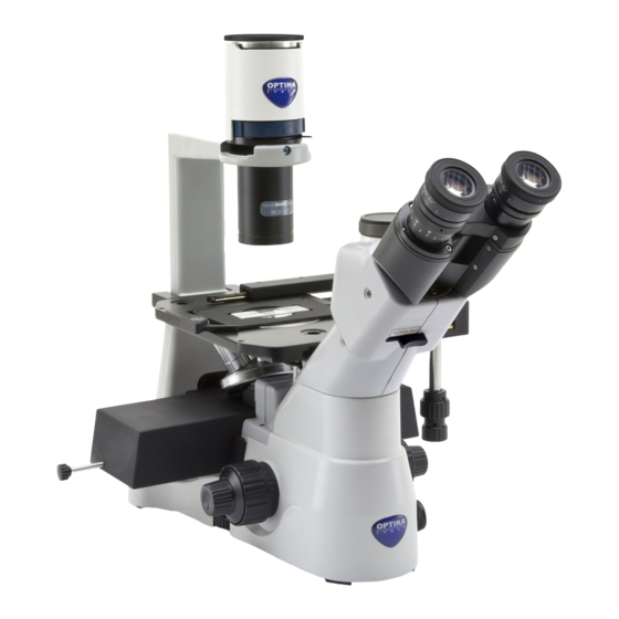
  • Seite 205: Beschreibung Des Instruments

    Beschreibung des Instruments IM-3 OKULARE GEHÄUSE FOTO/TV- AUSGANG FILTERTRÄGER PHASENKONTRAST- SCHIEBER KONDENSATOR DIOPTRIENVER- STELLUNGSRING TISCHPLATTE OBJEKTTISCH LICHTWE- GAUSWAHL OBJEKTIVE FEINTRIEBKNOPF HAUPTSCHALTER MIKROSKOPKÖRPER SPANNUNGSEIN- STELLUNGSRING GROBTRIEBKNOPF Seite 205...
  • Seite 206: Im-3F

    IM-3F FOTO/TV- AUSGANG LED GEHÄUSE OKULARE KONDENSATOR FILTERTRÄGER PHASENKONTRAST-SCHIEBER UV-SCHIRM DIOPTRIENVER- TISCHPLATTE STELLUNGSRING OBJEKTTISCH QUECKSILBER- LAMPGEHÄUSE LICHTWE- GAUSWAHL MIKROSKOPKÖRPER OBJEKTIVE FLUORESZENZ-WÜR- FELHEBEL FLUORESZENZ HAUPTSCHALTER NETZTEIL FEINTRIEBKNOPF SPANNUNGSEIN- STELLUNGSRING GROBTRIEBKNOPF Seite 206...
  • Seite 207: Im-3Fl4

    IM-3FL4 LED GEHÄUSE OKULARE KONDENSATOR FOTO/TV- AUSGANG FILTERTRÄGER DIOPTRIENVER- STELLUNGSRING UV-SCHIRM TISCHPLATTE LICHTWE- OBJEKTTISCH GAUSWAHL QUECKSILBER- LAMPGEHÄUSE FLUORESZENZ- FLUORESZENZ WÜRFELHEBEL NETZTEIL FEINTRIEBKNOPF MIKROSKOPKÖRPER DURCHLICHT EIN- GROBTRIEBKNOPF STELLKNOPF Seite 207...
  • Seite 208: Im-3Ld2

    IM-3LD2 LED GEHÄUSE FOTO/TV- AUSGANG OKULARE KONDENSATOR DIOPTRIENVER- STELLUNGSRING FILTERTRÄGER PHASENKONTRAST- SCHIEBER TISCHPLATTE OBJEKTTISCH LICHTWE- GAUSWAHL OBJEKTIVE FLUORESZENZ- AUFLICHT WÜRFELHEBEL EINSTELLKNOPF FEINTRIEBKNOPF DURCHLICHT EINSTELLKNOPF MIKROSKOPKÖRPER GROBTRIEBKNOPF Seite 208...
  • Seite 209: Im-3Ld4

    IM-3LD4 LED GEHÄUSE FOTO/TV- AUSGANG OKULARE KONDENSATOR DIOPTRIENVER- STELLUNGSRING FILTERTRÄGER TISCHPLATTE OBJEKTTISCH LICHTWE- GAUSWAHL OBJEKTIVE X-Y-BEWE- GUNGSKNÖPFE FLUORESZENZ- AUFLICHT WÜRFELHEBEL EINSTELLKNOPF FEINTRIEBKNOPF DURCHLICHT EINSTELLKNOPF MIKROSKOPKÖRPER GROBTRIEBKNOPF Seite 209...
  • Seite 210: Im-3Ld4D

    IM-3LD4D MONITOR KONDENSATOR FILTERTRÄGER USB KAMERA OKULARE DIOPTRIENVER- STELLUNGSRING TISCHPLATTE OBJEKTTISCH LICHTWE- GAUSWAHL OBJEKTIVE X-Y-BEWE- GUNGSKNÖPFE FLUORESZENZ- WÜRFELHEBEL AUFLICHT EINSTELLKNOPF FEINTRIEBKNOPF DURCHLICHT EINSTELLKNOPF MIKROSKOPKÖRPER GROBTRIEBKNOPF Seite 210...
  • Seite 211 IM-3LD4D (Rückseite) MONITOR-STROM- VERSORGUNGSAN- SCHLUSS HDMI-STECKER LED-STROMVER- MONITOR-STROMVER- SORGUNGSKABEL SORGUNGSKABEL USB-MOUSE / KEYBOARD- EMPFÄNGER PC STICK USB-KABEL (ZU KAMERA) PC STICK-STROMVER- SORGUNGSKABEL HDMI-KABEL (ZU MONITOR) Seite 211...
  • Seite 212: Öffnung Der Verpackung

    Öffnung der Verpackung Das Mikroskop ist in einem geformten Schaumpolystyrol Verpackung verpackt. Entfernen Sie das Klebeband von der Verpackung und ziehen Sie die obere Hälfte der Verpackung hoch. Beachten Sie bitte, die optischen Be- standteile (Objektive und Okulare) nicht fallen zu lassen oder nicht zu beschädigen. Ziehen Sie das Mikroskop aus der Verpackung mit beiden Händen (eine um den Arm und eine um die Basis) heraus und legen Sie es auf eine stabile Oberfläche.
  • Seite 213: Im-3F / Im-3Fl4

    IM-3F / IM-3FL4 ① ⑭ ② ⑮ ④ ⑨ ③ ⑩ ⑫ ⑬ ⑥ ⑤ ⑪ ⑯ ⑰ ⑱ ⑲ ⑦ ⑧ ① Mikroskop-Körper ⑬ Fluoreszenzbeleuchter ② Kondensator ⑭ Quecksilberlampgehäuse ③ LED-Leuchte ⑮ Mikroskop netzteile ④ Fluoreszenz netzteile + kabel ⑯ Phasenkontrast-Schieber ⑤ Stromkabel • Lieferung mit IM-3F ⑥ Filterhalter • Optional mit IM-3FL4 ⑦ Metalltischplatte ⑰ Zentrier-Teleskop ⑧ Glastischplatte • Lieferung mit IM-3F ⑨ Objektive • Optional mit IM-3FL4...
  • Seite 214: Im-3Ld2

    IM-3LD2 ③ ② ① ⑤ ⑭ ⑰ ⑦ ⑨ ⑯ ⑧ ⑩ ⑪ ④ ⑫ ⑮ ⑥ ⑬ ① Mikroskop-Körper ⑩ Okulare ② Kondensator ⑪ Objekttisch-Einsätze ③ LED-Leuchte ⑫ Zentrier-Teleskop ⑬ Grünfilter IF550 ④ Anti-Glühkappe ⑤ Mechanischer Tisch ⑭ Objektiv-Reinigungstuch ⑥ Phasenkontrast-Schieber ⑮ UV-Schirm ⑦ Glastischplatte ⑯ Staubschutzhaube...
  • Seite 215: Im-3Ld4

    IM-3LD4 ③ ② ① ⑨ ⑫ ⑭ ⑦ ④ ⑬ ⑧ ⑪ ⑤ ⑥ ⑩ ① Mikroskop-Körper ⑧ Metalltischplatte ② Kondensator ⑨ Mechanischer Tisch ③ LED-Leuchte ⑩ UV-Schirm ④ Objektive ⑪ Objekttisch-Einsätze ⑤ Okulare ⑫ Objektiv-Reinigungstuch ⑥ Anti-Glühkappe ⑬ Staubschutzhaube ⑦ Glastischplatte ⑭ Netzgerät + Netzkabel Seite 215...
  • Seite 216: Im-3Ld4D

    IM-3LD4D ③ ② ④ ⑨ ① ⑲ ⑪ ⑰ ⑦ ⑳ ⑧ ⑫ ⑤ ⑩ ⑥ ⑱ ⑬ ⑯ ⑭ ⑮ ① Mikroskop-Körper ⑪ Kamera ② Kondensator ⑫ “C” mount ③ LED-Leuchte ⑬ Maus ⑭ Tastatur ④ Anti-Glühkappe ⑤ Objektive ⑮ Monitor ⑥ Okulare ⑯ UV-Schirm ⑦ Glastischplatte...
  • Seite 217: Zusammenbau

    Zusammenbau Montage der Objektive ② 1. Drehen Sie den Großtriebknopf ① bis der Revolver sich in die tiefste Position befindet. • Aus Sicherheitsgründen wird der Revolver vor dem Versand in die tiefste Position gesetzt und der Spannungsring ② wird zur richtigen Spannung ein- gestellt.
  • Seite 218: Montage Der Tischplatte

    Montage der Tischplatte 1. Prüfen Sie, die Einlage perfekt horizontal ist, als der Glaseinsatz verwendet wird. 2. Setzen Sie den Einsatz in die Öffnung des Objekttisch ein. (Fig. 5) F ig. 5 ig. 5 Montage der Okulare Nehmen Sie den Verschluss aus den Okulartuben heraus, setzen Sie die Okulare in den Tuben ein.
  • Seite 219: Montage Des Led-Gehäuses

    Montage des LED-Gehäuses • Alle Modelle außer IM-3LD4D 1. Setzen Sie das Speisekabel in den Stecker ein. 2. Setzen Sie den Lampenträger in die Löcher der Leuchte Gruppe sehr vorsichtig ein. (Fig. 9) F ig. 9 ig. 9 • IM-3LD4D Die LED des IM-3LD4D ist bereits auf der Leuchte vorin- stalliert.
  • Seite 220: Installieren Des Pc-Bildschirms (Im-3Ld4D)

    Installieren des PC-Bildschirms (IM-3LD4D) • Für die Installation der Kamera lesen Sie bitte Kapitel 15.1. 1. Richten Sie die Kerbe ① mit der Halterung ② an der ① Beleuchtung aus. (Fig. 13) F ig. 13 ig. 13 ② 2. Wenn der Monitor vollständig eingesetzt ist, ziehen Sie das Rad ③...
  • Seite 221: Anschluss Der Netzteil

    6. Stecken Sie das Monitor-Netzteil in den Anschluss „F“ am Monitor. (Fig. 17) 7. Stecken Sie die andere Seite des HDMI-Kabels in den Anschluss „G“ am Monitor. (Fig. 17) F ig. 17 ig. 17 8. Stecken Sie das USB3.0-Kabel auf der Rückseite der Kamera ein.
  • Seite 222: Montage Der Fluoreszenz

    7.11 Montage der Fluoreszenz (Nur IM-3F und IM-3FL4) • Trennen Sie alle elektrischen Kabel, bevor Sie die Lampe installieren oder austauschen. • Die Lampe hat eine Anode und eine Kathode in verschiedenen Größen. Beachten Sie bei der Montage die Polaritäten unter Beachtung der Abmessungen des Leuchtenkopfes. •...
  • Seite 223 4. Entfernen einen Rändelknöpfe Filterhalteschlitten und setzen Sie den Schlitten in den Schlitz auf der Rückseite des Mikroskops ein. (Fig. 22) 5. Nach dem Einsetzen des Schlittens den Rändelknopf wieder einschrauben. F ig. 22 ig. 22 • IM-3F 6. Schrauben Sie die Klemme mit der eingravierten Be- schriftung G auf das Ende der Stange.
  • Seite 224 9. Öffnen Sie den Lampenkörper mit der Türklemmschraube ① und ziehen Sie den Lampenhalter heraus. (Fig. 26) ① F ig. 26 ig. 26 ② 10. Entfernen Kunststoffblock Lampenkörper (oder verwendete Lampe im Austauschfall), indem Sie die beiden ③ Verriegelungsschrauben ③ lösen. (Fig. 7) ②...
  • Seite 225 13. Stecken Sie das Netzkabel in den Anschluss. (Fig. 30) • Die Eingangsspannung des Vorschaltge- rätes beträgt 110-240Vac. • Bitte verwenden Sie das mitgelieferte Standardnetzkabel. Wählen Sie bei feh- lendem oder beschädigtem Kabel ein ge- eignetes Kabel aus. • Schließen Sie die Spannungsversorgung korrekt an, achten Sie auf eine gute Erd- verbindung.
  • Seite 226: Hellfeldbeobachtung (Durchlicht)

    Hellfeldbeobachtung (Durchlicht) (Verwendete Befehle) (Kapitel) Hauptschalter Schalten Sie den Hauptschalter auf “I” (ON) und stellen Sie die Lichtintensität ein Durchlicht einstellknopf Objekttisch Eine Zubereitung auf den Objekttisch. Revolver Setzen Sie die 10X objektiv in den optischen Pfad ein Fein- und großtriebdrehknopf Fokussierung der Vorbereitung Optischer kopf Einstellen des Augenabstandes...
  • Seite 227: Verwendung Des Mikroskops Im Hellfeld (Durchlicht)

    Verwendung des Mikroskops im Hellfeld (Durchlicht) Einschalten des Mikroskops Stellen Sie den Hauptschalter ① auf die Position “I” (ON). (Fig. 31) ① F ig. 31 ig. 31 Einstellen der Lichtintensität Verwenden Sie das Einstellrad für die Lichtintensität ②, um die Beleuchtungsspannung zu erhöhen oder zu verringern. (Fig.
  • Seite 228: Einstellung Des Augenabstandes

    Einstellung des Augenabstandes ① Beobachten Sie mit beiden Augen und unterstützen Sie die Gruppe der Okulare. Drehen Sie diese entlang der gemeinsamen Achse, bis Sie ein einziges Sichtfeld erhalten. • Abstufung Interpupillardistanzeigers ①, die durch den Punkt “.” Am Okularsockel gekennzeichnet ist, zeigt den Abstand zwischen den Augen des Operators.
  • Seite 229 IM-3 WAHLSCHALTER LEUCHTKRAFT APPLIKATIONEN Binokulare Beobachtung und Mikro- 50% bei binokularer Beobachtung / 50% fotografie können gleichzeitig durch- bei Mikrofotografie geführt werden 100% bei binokularer Beobachtung / 0% Binokulare Beobachtung bei Mikrofotografie IM-3F / IM-3FL4 / IM-3LD2 / IM-3LD4 / IM-3LD4D WAHLSCHALTER LEUCHTKRAFT APPLIKATIONEN...
  • Seite 230: Installieren Von Objekttisch-Einsätzer

    9.8.1 Installieren von Objekttisch-Einsätzer 1. Montieren Sie den Halter in den Mechanischer Tisch. (Fig. 40) F F ig. 40 ig. 40 2. Multiwell-Platten können direkt in den Mechanischer Tisch werden. (Fig. 41) F ig. 41 ig. 41 Aperturblende Der numerische Öffnungswert (A.N.) der Aperturblende beeinflusst den Kontrast des Bildes.
  • Seite 231: Verwendung Der Farbfilter

    9.10 Verwendung der Farbfilter Wählen Sie die Farbfilter nach Bedarf. (Fig. 44) FILTER ANWENDUNG Grün (IF550) Phasenkontrast Mikrosko- F ig. 44 ig. 44 Seite 231...
  • Seite 232: Verwendung Des Mikroskops Im Phasenkontrast (Durchlicht)

    10. Verwendung des Mikroskops im Phasenkontrast (Durchlicht) 10.1 Installieren von Phasenkontrast-Schieber 1. Setzen Sie den Schlitten in die Beleuchtungsanordnung ein, wobei der bedruckte Teil nach oben zeigt. (Fig. 45) 2. Bewegen Sie den Schlitten in die gewünschte Position, bis er mit einem Klick einrastet. 3.
  • Seite 233 4. Fokus Sie sich beim CT auf das Phasenringbild des Kondensators (Licht) ① und der Linse (dunkel) ②. ① Wenn das Bild des Lichtrings nicht scharf ist, stellen Sie den Drehmoment- und Winkelschlüssel ein, bis das Bild des Lichtrings scharf ist. (Fig. 48) 5.
  • Seite 234: Verwendung Des Mikroskops Im Rpc (Durchlicht)

    11. Verwendung des Mikroskops im RPC (Durchlicht) Der Relief-Phasenkontrast (RPC) ist eine Modifikation des herkömmlichen Phasenkontrasts, die zu sichtbaren Verbesserungen der Bildqualität in der optischen Mikroskopie führt. Insbesondere können die folgenden Parameter verbessert werden: Kontrast, Schärfentiefe, Schärfe, Dreidimensionalität, Ebenheit und Halo-Artefakte. Diese Effekte können erreicht werden, wenn die Phasenringe des Kondensors durch Spaltringe ersetzt werden.
  • Seite 235: Rpc Beobachtung

    11.3 RPC beobachtung • RPC-Ringe benötigen keine Zentrierung. 1. Legen Sie eine Probe auf den Objekttisch und fokus- sieren Siet. 2. Überprüfen Sie, dass der RPC ring und das Objektiv übereinanderstimmen und dass Beide stetig am Click- Stop eingestellt sin. 3.
  • Seite 236: Beobachtungsverfahren Im Fluoreszenz (Im-3F / Im-3Fl4)

    12. Beobachtungsverfahren im Fluoreszenz (IM-3F / IM-3FL4) (Verwendete Befehle) (Kapitel) Stellen Sie den Schalter der Stromversorgung auf “I” Fluoreszenz-Netzteile (ON) und Warten Sie, bis sich der Lichtbogen stabilisiert hat (5 oder 10 Minuten). Stellen Sie eine Zubereitung auf den Tisch. Objekttisch Setzen Sie den passenden Filterhalter in den optischen Fluoreszenz-Filterschieber...
  • Seite 237: Verwendung Des Mikroskops Im Fluoreszenz (Auflicht)

    14. Verwendung des Mikroskops im Fluoreszenz (Auflicht) 14.1 Zentrieren der HBO-Lampe Nur IM-3F / IM-3FL4 • Warten Sie etwa 5 Minuten, bevor Sie dies tun, damit sich die Lampe richtig aufwärmen kann. 1. Schalten Sie die Quecksilberdampflampe mit dem Netzschalter ① ein. (Fig. 55) 2. Drehen Sie den Revolver in eine leere Position ①...
  • Seite 238 6. Zentrierschrauben ③ an der Seite des Lampenkörpers verwenden, um das Bild des Lichtbogens zu zentrieren. (Fig. 59-60) F ig. 59 ig. 59 F ig. 60 ig. 60 7. Mit der Fokussierschraube der Sammellinse ② das Bild vergrößern, bis eine homogene Ausleuchtung erreicht ist.
  • Seite 239: Zentrieren Der Feldblende

    14.2 Zentrieren der Feldblende ② ① Nur IM-3F / IM-3FL4 1. Legen Sie die Probe auf den Couchtisch, setzen Sie die 10x-Objektiv in den Strahlengang ein und fokussieren Sie auf. 2. Drehen Sie den Feldblendenhebel ①, um die Membran vollständig zu schließen. (Fig. 63) 3.
  • Seite 240: Verfügbare Fluoreszenzfilterwürfel

    • IM-3FL4 Bewegen Sie den Wählhebel (links am Mikroskop) ④, um den gewünschten Filter einzusetzen: B, G (V und UV - optional). (Fig. 67) ⑤ F ig. 67 ig. 67 • IM-3LD2 / IM-3LD4 / IM-3LD4D Bewegen Sie den Filterhebel (auf der linken Seite des Mikroskops) ⑥, um den gewünschten Filterwürfel einzusetzen (siehe Tabellen unten).
  • Seite 241: Installation Des Fluoreszenzfilters

    • IM-3LD4 / IM-3LD4D D I C H R O I - F I LT E R ANREGUNGS- EMISSIONSFIL- T I S C H E R ANWENDUNG NAME FILTER SPIEGEL • DAPI M-1233 325-375 nm 415 nm 435LP nm • Hoechst •...
  • Seite 242: Verwendung Der Anti-Glühkappe

    6. Montieren Sie den Filterschieber wieder am Mikroskop. 7. Schließen Sie die seitliche Abdeckung. 8. Bringen Sie den Haftmarker ⑦ für den Fluoreszenz- würfel auf der Seitenabdeckung an. (Fig. 71) 9. Schließen Sie die Spannungsversorgung an. 10. Beginn der Arbeit. ⑦...
  • Seite 243: Verwendung Des Filterhalter-Schiebers

    14.6.2 Verwendung des Filterhalter-Schiebers • Bewegen Sie den Schlitten nach rechts oder links, um die gewünschte Position einzugeben. Ein Klick bestätigt die korrekte Position des Schlittens. 1. Durch Eingabe der Position ① (sofern ein ND-Filter installiert ist) wird das aus dem Lampengehäuse kommende Licht je nach Filtertyp reduziert.
  • Seite 244: Gleichzeitiger Phasenkontrast + Fluoreszenzanwendung

    15. Gleichzeitiger Phasenkontrast + Fluoreszenzanwendung • Fluoreszenzmodelle ermöglichen die Beobachtung im Durchlicht Phasenkontrast in Kombination mit Auflicht Fluoreszenz. Schnell zerfallende Proben sollten zunächst in der Fluoreszenz und dann im Phasenkontrast beobachtet werden. Kombinierte Beobachtungen erleichtern die Identifizierung bestimmter Bereiche der Probe, die Fluoreszenz emittieren. 15.1 IM-3F / IM-3FL4 1. Schalten Sie die Stromversorgung für die HBO-Leuchtstofflampe ein und warten Sie 5 Minuten, bevor sich der Lichtbogen stabilisiert.
  • Seite 245: Mikrofotografie

    16. Mikrofotografie 16.1 Verwendung von C-Mount Kameras ② 1. Lösen Sie die Sicherungsschraube ① am Binokulartubus und entfernen Sie die Staubkappe ②. (Fig. 75) ① F ig. 75 ig. 75 2. Schrauben Sie den Adapterschritt “C” ③ an die Kamera ④...
  • Seite 246: Wartung

    Montieren Sie die Objektive und Okulare nicht ab, um sie zu reinigen. Am Besten verwenden Sie das OPTIKA Reinigungskit (siehe Katalog) Falls das Mikroskop aus Wartungszwecken an Optika zurückgeschickt werden muss, verwenden Sie bitte im- mer die Originalverpackung. Seite 246...
  • Seite 247: Probleme Und Lösungen

    18. Probleme und Lösungen Lesen Sie die Informationen in der folgenden Tabelle, um Probleme bei der Bedienung zu behebeni. PROBLEM URSACHE LÖSUNG I. Optisches System: Der LED ist eingeschaltet, aber das Der Stecker des LED-Gehäuses ist Verbinden Sie das LED-Gehäuse mit Sichtfeld ist dunkel.
  • Seite 248 II. Mechanischer System: Der makrometrische Knopf ist schwer Einstellring zu fest spannen Lösen Sie den Einstellring für die zu drehen. Spannung. Die Fokussierung ist instabil. Einstellring zu locker gespannt Ziehen Sie den Einstellring für die Spannung an. III. Elektrischer System: Die LED leuchtet nicht.
  • Seite 249: Wiederverwertung

    Wiederverwertung Gemäß dem Artikel 13 vom Dekret Nr. 151 vom 25.07.2005 “Umsetzung der Richtlinien 2002/95/EG, 2002/96/ EG und 2003/108/EG in Bezug auf die Verwendung gefährlicher Stoffe in elektrischen und elektronischen Gerä- ten sowie die Abfallentsorgung”. Das Symbol vom Müllcontainer erscheint auf dem Gerät oder der Verpackung und weist darauf hin, dass das Produkt Ende des Lebens separat von anderen Abfällen entsorgt werden muss.
  • Seite 250 OPTIKA S.r.l. ® Via Rigla, 30 - 24010 Ponteranica (BG) - ITALY Tel.: +39 035.571.392 info@optikamicroscopes.com - www.optikamicroscopes.com OPTIKA Spain spain@optikamicroscopes.com OPTIKA USA usa@optikamicroscopes.com OPTIKA China china@optikamicroscopes.com OPTIKA India india@optikamicroscopes.com OPTIKA Central America camerica@optikamicroscopes.com...
  • Seite 251 Série IM MANUAL DE INSTRUÇÕES Modelo IM-3 IM-3F IM-3FL4 IM-3LD2 IM-3LD4 IM-3LD4D Ver. 5.2 2021...
  • Seite 252 Tabela de Conteúdos Advertência Símbolos Informações sobre a segurança Utilização prevista Descrição do instrumento IM-3 IM-3F IM-3FL4 IM-3LD2 IM-3LD4 IM-3LD4D Desembalando IM-3 IM-3F / IM-3FL4 IM-3LD2 IM-3LD4 IM-3LD4D Procedimento de instalação Montagem dos objectivos Montagem de extensão lateral e platina mecânica Montagem do inserto Instalação do ocular Instalação do condensador...
  • Seite 253 14.5 Uso da capa anti-reflexo 14.6 Suporte do filtro / Obturador 14.6.1 Inserir um filtro ND 14.6.2 Uso do suporte do filtro 15. Observação simultânea Contraste de fase + Fluorescência 15.1 IM-3F / IM-3FL4 15.2 IM-3LD2 / IM-3LD4 / IM-3LD4D 16. Microfotografia 16.1 Uso de câmaras de paso “C” 16.2 Uso de câmaras Reflex 17. Manutenção 18. Resolução de problemas Eliminação Página 253...
  • Seite 254: Informações Sobre A Segurança

    Advertência Este microscópio é um instrumento científico de alta precisão, projectado para durar um longo tempo com manutenção mínima; a sua realização respeita os melhores padrões ópticos e mecânicos, para que possa ser utilizado diariamente. Recordamos que este manual contém informações importantes para a segurança e a manutenção do instrumento, portanto deve ser colocado à...
  • Seite 255: Descrição Do Instrumento

    Descrição do instrumento IM-3 OCULAR COMPARTIMENTO SAÍDA DE FOTO/TV SUPORTE DO FILTRO SLIDE DE CONTRASTE DE FASE CONDENSADOR ANEL DE PLACA DA AJUSTE PLATINA DIOPTRICO PLATINA SELECTOR DO CAMINHO DA OBJETIVAS BOTAO DE AJUSTE MICROMÉTRICO INTERRUPTOR PRINCIPAL BASE DE MICROSCÓPIO ARO DE AJUSTE DE TENSAO...
  • Seite 256: Im-3F

    IM-3F SAÍDA DE FOTO/TV COMPARTIMENTO OCULAR CONDENSADOR SUPORTE DO FILTRO SLIDE DE CONTRASTE DE FASE ECRÃ UV ANEL DE AJUSTE PLACA DA PLATINA DIOPTRICO PLATINA CORPO DA LÂMPADA FLUORESCENTE SELECTOR DO CAMINHO DA BASE DE OBJETIVAS MICROSCÓPIO ALAVANCA DO CUBO DE FLUORESCÊNCIA ALIMENTAÇÃO POR INTERRUPTOR...
  • Seite 257: Im-3Fl4

    IM-3FL4 COMPARTIMENTO OCULAR CONDENSADOR SAÍDA DE FOTO/TV SUPORTE DO FILTRO ANEL DE AJUSTE DIOPTRICO ECRÃ UV PLACA DA PLATINA SELECTOR DO PLATINA CAMINHO DA CORPO DA LÂMPADA FLUORESCENTE ALAVANCA DO CUBO ALIMENTAÇÃO POR DE FLUORESCÊNCIA FLUORESCÊNCIA BOTAO DE AJUSTE MICROMÉTRICO BASE DE MICROSCÓPIO BOTÃO DE...
  • Seite 258: Im-3Ld2

    IM-3LD2 COMPARTIMENTO SAÍDA DE FOTO/TV OCULAR CONDENSADOR ANEL DE AJUSTE DIOPTRICO SUPORTE DO FILTRO SLIDE DE CONTRASTE DE FASE PLACA DA PLATINA PLATINA SELECTOR DO CAMINHO DA OBJETIVAS ALAVANCA DO CUBO DE BOTÃO DE FLUORESCÊNCIA AJUSTAMENTO DA LUZ REFLETIDA BOTAO DE AJUSTE MICROMÉTRICO BOTÃO DE AJUSTAMENTO DA...
  • Seite 259: Im-3Ld4

    IM-3LD4 COMPARTIMENTO SAÍDA DE FOTO/TV OCULAR CONDENSADOR ANEL DE AJUSTE DIOPTRICO SUPORTE DO FILTRO PLACA DA PLATINA PLATINA SELECTOR DO CAMINHO DA OBJETIVAS BOTÕES DE MOVIMENTO X-Y ALAVANCA DO CUBO DE BOTÃO DE FLUORESCÊNCIA AJUSTAMENTO DA LUZ REFLETIDA BOTAO DE AJUSTE MICROMÉTRICO BOTÃO DE AJUSTAMENTO DA...
  • Seite 260: Im-3Ld4D

    IM-3LD4D MONITOR CONDENSADOR SUPORTE DO FILTRO CÂMARA USB OCULAR ANEL DE AJUSTE DIOPTRICO PLACA DA PLATINA PLATINA SELECTOR DO CAMINHO DA OBJETIVAS BOTÕES DE MOVIMENTO X-Y ALAVANCA DO CUBO DE FLUORESCÊNCIA BOTÃO DE AJUSTAMENTO DA LUZ REFLETIDA BOTAO DE AJUSTE MICROMÉTRICO BOTÃO DE AJUSTAMENTO DA...
  • Seite 261 IM-3LD4D (lado de trás) CONECTOR CABO DE CONECTOR HDMI ALIMENTAÇÃO MONITOR CABO DE CABO DE ALIMENTAÇÃO ALIMENTAÇÃO MONITOR RECEPTOR USB RATO / TECLADO PC STICK CABO USB (PARA CÂMARA) CABO DE ALIMENTAÇÃO PC STICK CABO HDMI (PARA MONITOR) Página 261...
  • Seite 262: Desembalando

    Desembalando O microscópio é alojado em um recipiente de isopor moldado. Remova a fita da borda do recipiente e levante a metade superior do recipiente. Tome algum cuidado para evitar que os itens ópticos (objetivas e oculares) cair e ficar danificado. Usando ambas as mãos (uma ao redor do braço e outra ao redor da base), levante o micros- cópio do recipiente e coloque-o em uma mesa estável.
  • Seite 263: Im-3F / Im-3Fl4

    IM-3F / IM-3FL4 ① ⑭ ② ⑮ ④ ⑨ ③ ⑩ ⑫ ⑬ ⑥ ⑤ ⑪ ⑯ ⑰ ⑱ ⑲ ⑦ ⑧ ① Microscópio ⑬ Iluminador de Fluorescência ② Condensador ⑭ Corpo da lâmpada HBO ③ Compartimento LED ⑮ Transformador microscópio ④ Alimentação por fluorescência + cabo ⑯ Slide de contraste de fase ⑤ Cabo de ligação de fluorescência • F ornecido com IM-3F ⑥ Soporte para filtro • O pcional com IM-3FL4...
  • Seite 264: Im-3Ld2

    IM-3LD2 ③ ② ① ⑤ ⑭ ⑰ ⑦ ⑨ ⑯ ⑧ ⑩ ⑪ ④ ⑫ ⑮ ⑥ ⑬ ① Microscópio ⑩ Oculares ② Condensador ⑪ Insertos da platina ③ Compartimento LED ⑫ Telescópio de centragem ④ Capa anti-reflexo ⑬ Filtro verde IF550 ⑤ Platina mecânica ⑭ Lenços de limpeza de lentes ⑥ Slide de contraste de fase ⑮ Ecrã UV ⑦ Inserto de vidrio ⑯ Capa de pó...
  • Seite 265: Im-3Ld4

    IM-3LD4 ③ ② ① ⑨ ⑫ ⑭ ⑦ ④ ⑬ ⑧ ⑪ ⑤ ⑥ ⑩ ① Microscópio ⑧ Inserto de metal ② Condensador ⑨ Platina mecânica ③ Compartimento LED ⑩ Ecrã UV ④ Objetivas ⑪ Insertos da platina ⑤ Oculares ⑫ Lenços de limpeza de lentes ⑥ Capa anti-reflexo ⑬ Capa de pó ⑦ Inserto de vidrio ⑭ Fonte de alimentação + cabo de alimentação Página 265...
  • Seite 266: Im-3Ld4D

    IM-3LD4D ③ ② ④ ⑨ ① ⑲ ⑪ ⑰ ⑦ ⑳ ⑧ ⑫ ⑤ ⑩ ⑥ ⑱ ⑬ ⑯ ⑭ ⑮ ① Microscópio ⑪ Câmara ② Condensador ⑫ Paso “C” ③ Compartimento LED ⑬ Rato ④ Capa anti-reflexo ⑭ Teclado ⑤ Objetivas ⑮ Monitor ⑥ Oculares ⑯ Ecrã UV ⑦ Inserto de vidrio...
  • Seite 267: Procedimento De Instalação

    Procedimento de instalação Montagem dos objectivos ② 1. Rodar o manípulo de regulação macrométrica ① até que a torre de objetivas esteja na posição mais baixa. • Para garantir a segurança durante o transporte, antes da expedição a torre é colocada na posição mais baixa e o anel de regulação da tensão ②...
  • Seite 268: Montagem Do Inserto

    Montagem do inserto 1. Ao usar o elemento de vidro, certifique-se de que a in- serção esteja na horizontal. 2. Inserir o inserto na abertura da platina. (Fig. 5) F ig. 5 ig. 5 Instalação do ocular Remova a tampa dos tubos do ocular e insira as oculares nos tubos.
  • Seite 269: Instalação Do Compartimento Led

    Instalação do compartimento LED • Todos os modelos excepto IM-3LD4D 1. Insira o cabo de alimentação na ficha da tomada. 2. Pressione cuidadosamente o suporte do LED nos orifí- cios do sistema de iluminação. (Fig. 9) F ig. 9 ig. 9 •...
  • Seite 270: Instalação Do Monitor Pc (Im-3Ld4D)

    Instalação do monitor PC (IM-3LD4D) • Para a instalação da câmara, consulte por favor o ca- pítulo 15.11. 1. Alinhar o entalhe ① com o suporte ② no iluminador. ① (Fig. 13) F ig. 13 ig. 13 ② 2. Quando o monitor estiver totalmente inserido, aperte a roda ③...
  • Seite 271: Conexão Da Fonte De Alimentação

    6. Ligar a fonte de alimentação do monitor no conector “F” do monitor. (Fig. 17) 7. Ligar o outro lado do cabo HDMI no conector “G” do monitor. (Fig. 17) F ig. 17 ig. 17 8. Ligar o cabo USB3.0 na parte de trás da câmara. (Fig. F ig.
  • Seite 272: Montagem De Fluorescência

    7.11 Montagem de fluorescência (Apenas IM-3F e IM3FL4) • Desligue todos os cabos eléctricos antes de instalar ou substituir a lâmpada. • A lâmpada tem um ânodo e um cátodo de tamanhos diferentes. Respeite a polaridade durante a montagem, respeitando as dimensões da lâmpada. •...
  • Seite 273 4. Remova um dos botões serrilhados do suporte do filtro e insira o botão na ranhura na parte de trás do microscópio. (Fig. 22) 5. Depois de inserir a corrediça, aparafuse novamente o botão serrilhado. F ig. 22 ig. 22 •...
  • Seite 274 9. Desaperte completamente o parafuso ① e remova o suporte da lâmpada. (Fig. 26) ① F ig. 26 ig. 26 10. Retire o bloco de plástico ② do suporte da lâmpada (ou da lâmpada esgotada em caso de substituição) desapertando os dois parafusos de bloqueio ③. (Fig. ③...
  • Seite 275 13. Conecte o cabo de alimentação à parte traseira da fon- te de luz. (Fig. 30) • A fonte de luz fluorescente opera com uma tensão de 110 a 240Va. • Use o cabo fornecido com o microscó- pio. Em caso de perda ou quebra, certi- fique-se de que o novo cabo é o mesmo que o fornecido.
  • Seite 276: Observação Em Campo Claro (Luz Transmitida)

    Observação em campo claro (luz transmitida) (Comandos utilizados) (Capìtulo) Ajuste a chave principal para “I” (ON) e ajuste a intensidade Interruptor principal da luz. Mostrador de intensidade de luz Coloque a amostra/preparação na platina Platina Insira a objetiva 10x no caminho da luz Revólver Focalize o espécime Botões de foco macro e micro...
  • Seite 277: Uso Do Microscópio Em Campo Claro (Luz Transmitida)

    Uso do microscópio em campo claro (luz transmitida) Ligar o microscópio Coloque o interruptor principal ① na posição “I” (ON). (Fig. ① F ig. 31 ig. 31 Ajuste da intensidade da luz Utilize a roda de ajuste da intensidade da luz ② para au- mentar ou diminuir a tensão de iluminação.
  • Seite 278: Uso De Ilhós De Borracha

    Ajustar a distância interpupilar ① Observando com ambos os olhos, segurar o grupo de oculares. Rodá-lo ao longo do eixo comum até obter um único campo visual. • A escala graduada no indicador de distância interpupilar ①, indicada pelo ponto “.” no suporte da ocular, mostra a distância interpupilar do operador.
  • Seite 279: Platina Mecânica E Porta-Preparação

    IM-3 ALAVANCA SELECTORA LUMINOSIDADE APLICAÇÕES A observação binocular e a micro- 50% para observação binocular / 50% fotografia podem ser realizadas para microfotografia simultaneamente 100% para observação binocular / 0% Observação binocular para microfotografia IM-3F / IM-3FL4 / IM-3LD2 / IM-3LD4 / IM-3LD4D ALAVANCA SELECTORA LUMINOSIDADE APLICAÇÕES...
  • Seite 280: Instalação De Insertos Da Platina

    9.8.1 Instalação de insertos da platina 1. Instalar o suporte na platina mecânica. (Fig. 40) F F ig. 40 ig. 40 2. Placas de múltiplos poços podem ser directamente in- seridas na platina mecânica. (Fig. 41) F ig. 41 ig. 41 Diafragma de abertura O valor de abertura numérica (N.A.) do diafragma de abertura afecta o contraste da imagem.
  • Seite 281: Uso De Filtros Coloridos

    9.10 Uso de filtros coloridos Escolha os filtros coloridos de acordo com as suas neces- sidades. (Fig. 33) FILTRO Verde (IF550) Microscopia de contraste de fase F ig. 44 ig. 44 Página 281...
  • Seite 282: Uso Do Microscópio Em Contraste De Fase (Luz Transmitida)

    10. Uso do microscópio em contraste de fase (luz transmitida) 10.1 Instalação da slide para contraste de fase 1. Insira a slide no sistema de iluminação com o lado im- presso virado para cima. (Fig. 45) 2. Mova a slide para a posição desejada até que ele trave com um clique.
  • Seite 283 condensador (claro) ① e na objetiva presente (escuro) ②. Se a imagem do anel de luz não estiver nítida, ajuste ① o CT até que a imagem do anel de luz fique nítida. (Fig. 5. Aperte os parafusos dos dois furos de centragem do slide por contraste de fase com as porcas extravasa- das fornecidas até...
  • Seite 284: Uso Do Microscópio Em Rpc (Luz Transmitida)

    11. Uso do microscópio em RPC (luz transmitida) O contraste de fase de alívio (RPC) é uma modificação do contraste de fase convencional que conduz a melhorias visíveis na qualidade da imagem em microscopia óptica. Especificamente, os seguintes parâmetros podem ser melhorados: contraste, profundidade focal, nitidez, tridimensionalidade, planicidade, e artefactos de auréola.
  • Seite 285: Observação Em Rpc

    11.3 Observação em RPC • Os anéis RPC não precisam de uma centragem. 1. Coloque um espécime na platina e focalize-o. 2. Verificar se o anel de RPC e a objetiva correspondem e se ambos estão fixos na posição de bloqueio.p. 3.
  • Seite 286: Observação Em Fluorescência (Im-3F / Im-3Fl4)

    12. Observação em fluorescência (IM-3F / IM-3FL4) (Comandos utilizados) (Capìtulo) Ligue a fonte de alimentação, posicione o botão de Fonte de luz fluorescente alimentação em “I” (ON) e aguarde 5-10 minutos para que a lâmpada estabilize. Coloque a amostra/preparação na platina Platina Insira o filtro no módulo de fluorescência Módulo de filtro de fluorescência correspondente à...
  • Seite 287: Utilização Do Microscópio Em Fluorescência (Luz Reflectida)

    14. Utilização do microscópio em fluorescência (luz reflectida) 14.1 Centragem da lâmpada HBO Apenas IM-3F / IM-3FL4 • Aguarde cerca de 5 minutos antes de prosseguir com esta operação para permitir que a lâmpada aqueça adequadamente. 1. Ligar a lâmpada de mercúrio accionando o interruptor da fonte de alimentação ①. (Fig. 55) ①...
  • Seite 288 6. Usando os parafusos de centralização ③ no lado do alojamento da lâmpada, centralize a imagem do arco. (Fig. 59-60) F ig. 59 ig. 59 F ig. 60 ig. 60 7. Usando o parafuso de focagem da lente do colector ②, amplie a imagem até...
  • Seite 289: Centragem Do Diafragma De Campo

    14.2 Centragem do diafragma de campo ① ② Apenas IM-3F / IM-3FL4 1. Coloque a amostra na platina, insira a lente de 10x no caminho óptico e focalize. 2. Gire a alavanca do diafragma de campo ① para fechar completamente o diafragma. (Fig. 63) 3.
  • Seite 290: Cubos De Filtro De Fluorescência Disponíveis

    • IM-3FL4 Mova a alavanca selectora (à esquerda do microscópio) ④ para inserir o filtro desejado: B, G (V e UV - opcional). (Fig. 67) ⑤ F ig. 67 ig. 67 • IM-3LD2 / IM-3LD4 / IM-3LD4D Mova a alavanca selectora (à esquerda do microscópio) ⑥ para inserir o filtro desejado (ver tabelas abaixo).
  • Seite 291: Instalação De Filtro De Fluorescência

    • IM-3LD4 / IM-3LD4D N O M E FILTRO DE ESPELHO FILTRO APLICAÇÕES FILTRO EXCITAÇÃO DICRÓICO EMISSÃO • DAPI M-1233 325-375 nm 415 nm 435LP nm • Hoechst • Coumarin M-1232 390-420 nm 440 nm 450LP nm • Pacific Blue •...
  • Seite 292: Inserir Um Filtro Nd

    6. Reinstalar o selector do filtro no microscópio. 7. Fechar a tampa lateral. 8. Aplicar o marcador adesivo ⑦ para o cubo de fluores- cência na tampa lateral. (Fig. 71) 9. Ligar a fonte de alimentação. 10. Comece a trabalhar. ⑦...
  • Seite 293 14.6.2 Uso do suporte do filtro • Mova o slide para a direita ou para a esquerda para inserir a posição desejada. Um clique confirmará a posição correta do slide. 1. Ao entrar na posição ① (sempre que um filtro ND é instalado) a luz proveniente do alojamento da lâmpada será...
  • Seite 294 15. Observação simultânea Contraste de fase + Fluorescência • Os modelos em fluorescência permitem a observação em luz transmitida Contraste de fase em combinação com luz reflectida para Fluorescência. Amostras com rápida perda de condições devem ser observadas primeiro na Fluorescência e depois no Contraste de Fase. A observação combinada permite identificar facilmente algumas áreas da amostra que emitem fluorescência. 15.1 IM-3F / IM-3FL4 1.
  • Seite 295 16. Microfotografia 16.1 Uso de câmaras de paso “C” ② 1. Desaperte o parafuso de aperto ① na porta trinocular e retire a tampa do pó ②. (Fig. 75) ① F ig. 75 ig. 75 2. Aparafuse o adaptador C-mount ③ à câmara ④ e insira o encaixe redondo do C-mount no orifício vazio da porta trinocular, depois aperte o parafuso de aperto ④...
  • Seite 296: Manutenção

    Não desmontar as objetivas ou os oculares para tentar limpá-los. Para um melhor resultado utilizar o kit de limpeza OPTIKA (ver catálogo). Se for necessário enviar o microscópio ao fabricante para a sua manutenção, pede-se que seja utilizada a embala- gem original.
  • Seite 297: Resolução De Problemas

    18. Resolução de problemas Reveja a informação na tabela abaixo para tentar solucionar problemas de operação. PROBLEMA CAUSA SOLUÇÃO I. Secção Óptica: O LED funciona, mas o campo de vi- O plugue do suporte da lâmpada não Conecte-os são permanece escuro. está...
  • Seite 298 II. Secção Mecânica O botão do foco macro está difícil de O anel de ajuste da tensão está muito Solte o anel de ajuste da tensão rodar apertado O foco é instável O anel do ajuste da tensão está muito Aperte o anel de ajuste da tensão solto IV.
  • Seite 299 Eliminação Art.13 Dlsg 25 de Julho de 2005 N°151. “De acordo com as Directivas 2002/95/CE, 2002/96/CE e 2003/108/CE relativas à redução do uso de substâncias perigosas em equipamentos eléctricos e electrónicos e à eliminação de resíduos. O símbolo do cesto no equipamento ou na sua caixa indica que o produto no final da sua vida útil deve ser recolhido separadamente dos outros resíduos.
  • Seite 300 Distribuito da: Geass s.r.l Via Ambrosini 8/2 - Torino Telefono: 011.22.91.578 Email:info@geass.com Sito:www.geass.com...

Diese Anleitung auch für:

Im-3Im-3fIm-3fl4Im-3ld2Im-3ld4Im-3ld4d

Inhaltsverzeichnis